Research Article
Survival and Prognostic Factors of Patients with Advanced CY1/P0 Gastric Cancer
Aoyagi K*, Kizaki J, Isobe T, Minami T and Akagi Y
Department of Surgery, Kurume University School of Medicine, Japan
*Corresponding author: Keishiro Aoyagi, Department of Surgery, Kurume University School of Medicine, 67 Asahi-machi, Kurume 830-0011, Japan
Published: 12 Sep, 2016
Cite this article as: Aoyagi K, Kizaki J, Isobe T, Minami T, Akagi Y. Survival and Prognostic Factors of Patients with Advanced CY1/ P0 Gastric Cancer. Clin Surg. 2016; 1: 1110.
Abstract
Introduction: The prognosis is poor for patients with gastric cancer with positive cytologic examinations of peritoneal lavage fluid (CY1), and optimal treatment is not established. Further, the surgical strategy for patients with free cancer cells in the intraperitoneal fluid in the absence of overt peritoneal metastasis (CY1/P0) is controversial. Here, we investigated the clinical outcomes and prognostic factors of patients with CY1/P0 gastric cancer.
Methods: We conducted an analysis of 39 patients with CY1/P0 gastric cancer who underwent gastrectomy and analyzed the clinical characteristics of patients who survived ≥3 years after surgery.
Results: Large type-3 and type-4 tumors, scirrhous (sci), and infiltrative growth cancers (INFc) were
associated with high recurrence rates. INFc was an independent risk factor for recurrence. Univariate analysis of overall survival (OS) showed that large type-3 and type-4 cancers, INFc, and lack of postoperative chemotherapy correlated with poor OS. Multivariate analysis showed that INFc and postoperative chemotherapy were independent prognostic factors. Univariate analysis showed that large type-3 and type-4 tumors, sci, INFc, and large tumor size correlated with poor progressionfree survival (PFS). Multivariate analysis showed that INFc was an independent prognostic factor of PFS. The numbers of long-term survivors with large type-3 and type-4 cancers were significantly lower compared with those with types 1–3.
Conclusion: In patients with CY1/P0 gastric cancer, INF and postoperative chemotherapy were independent prognostic factors of OS. The prognosis of large type-3 and type-4 P0/CY1-gastric cancers is very poor. Therefore, such patients should be treated with multidisciplinary therapy such as neoadjuvant chemotherapy.
Introduction
Gastric cancer is the fifth most common malignancy and the third leading cause of cancerrelated
death worldwide [1]. Patients with advanced gastric cancer have poor survival despite progress in multidisciplinary therapy [2,3]. Peritoneal dissemination (P1) is the most common metastatic pattern in gastric cancer, and the prognosis is extremely poor [4,5]. Patients with free intraperitoneal cancer cells without overt peritoneal metastasis (CY1/P0) are categorized as stage IV [6,7], and their 5-year overall survival (OS) rate after potentially curative surgery is 0–35% [8]. Peritoneal metastases arise from implantation of free peritoneal cancer cells, and CY1/P0 disease is considered a precursor of peritoneal metastasis. A positive cytologic examination of peritoneal lavage fluid (CY1) is considered a poor prognostic factor for patients with gastric cancer [9,10].
CY1 is therefore considered equivalent to distant metastasis, regardless of the presence of
peritoneal metastasis. Since 1999, CY1 gastric cancer has been considered stage IV disease according
to the Japanese Classification of Gastric Cancer (JCGC) stages [11]. Because peritoneal lavage
cytology is available in at least 90% of hospitals in Japan, this test may have the same prognostic
significance as peritoneal dissemination [12]. Moreover, the Union for International Cancer
Control (UICC) has considered CY1-gastric cancer as stage IV disease since 2009 [7]. Although
the prognosis of CY1 patients is poor, the optimal treatment for these patients is not established.
Therefore, the aim of the current study was to identify prognostic factors by evaluating the most
recent clinical outcomes of patients with CY1/P0 gastric cancer.
Materials and Methods
Patients
Between 2000 and 2014, 1830 patients with histologically confirmed primary gastric cancer underwent surgery at the Department of Surgery, Kurume University School of Medicine, Kurume, Japan. Peritoneal lavage cytology was principally performed for patients with tumors clinically invading the muscularispropria (cT2) or beyond, while it was omitted for tumors clinically confined to the mucosa or submucosa (cT1). Therefore, 684/1830 (37.4%) without distant metastasis underwent peritoneal lavage cytology, and 39 (5.7%) with cT2 or beyond were diagnosed as CY1/P0.
Clinicopathological factors
Analysis of tumor recurrence and prognosis were performed to
identify independent factors to predict recurrence and prognosis of the
39 patients with CY1/P0 gastric cancer who underwent gastrectomy.
Nineteen clinicopathological parameters were investigated, as listed
in (Table 1), including neoadjuvant chemotherapy, postoperative
chemotherapy, postoperative complications, and type of recurrence.
Tumor location, cross-sections of the stomach, macroscopic type,
and histology, depth of tumor invasion, stromal volume, tumor
infiltrative pattern (INF), lymphatic invasion (ly), venous invasion (v),
and lymph node metastasis (N) were evaluated according to the 3rd
English edition of the Japanese Classification of Gastric Carcinoma
[6]. The types of gastrectomy and lymph node dissection (D) were
evaluated according to the Japanese Gastric Cancer Treatment
Guidelines 2010 (ver. 3) [13]. In the present study, macroscopic type-
3 cancer (≥80 mm) was defined as large type-3 cancer.
Long-term survival
Patient’s alive ≥3 years after surgery are called long-term
survivors.
Statistical analyses
The clinical variables of patients with recurrence and long-term
survivors were compared using univariate analysis and the χ2 test.
We used a logistic regression method to extract significant factors
further analysis. OS and progression-free survival (PFS) rates were
calculated according to the Kaplan–Meier method, and differences
were evaluated using the log-rank test. Cox’s proportional hazards
regression model was used to identify prognostic factors for survival.
P< 0.05 was considered statistically significant. All statistical analyses
were performed using SPSS II software (IBM Co., Armonk, NY).
Table 1

Table 1
Patient characteristics (n = 39).
Table 2
Table 2
Characteristics of patients with and without recurrence.
Table 3
Table 3
Univariate and multivariate analysis of prognostic factors associated with overall survival (OS) and progression-free survival (PFS).
Figure 1
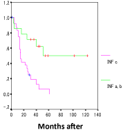
Figure 1
Kaplan–Meier analysis of overall survival (OS) of patients with
INFa,b and INFc . There was a significant difference in OS between the two
groups (P= 0.0011).
Results
Patients’ characteristics
The clinicopathological characteristics of 39 patients with CY1/P0
gastric cancer are presented in (Table 1). The median age was 73 years
(range, 30-86 years) and 24 patients were male. Thirty-three patients
(84.6%) had macroscopic infiltrating types that included type-3 and
type-4 tumors. There were 26 patients (66.7%) with large type-3 and
type-4 tumors. Twenty patients (51.3%) had scirrhous type (sci) and
25 (64.1) had infiltrative growth (INFc). The four (10.2%) patients
with type-4 cancer who underwent neoadjuvant chemotherapy were
administered S-1 + cisplatin (CDDP). Twenty-nine (74.4%) patients
received S-1or S-1 + another drug. Eleven (28.2%) patients required
second-line chemotherapy, and five (12.8%) patients required thirdline
or additional chemotherapy after surgery. Disease recurrence in
the peritoneum alone or with recurrence at another site was present
in 21 (53.8%) patients.
Recurrence and clinicopathological characteristics
Tumors recurred in 27 patients (69.2%). Univariate analysis
indicated a significant association between recurrence and
macroscopic type, stromal volume, and INF (Table 2). Multivariate
analysis showed that recurrence significantly associated with only
INF (hazard ratio [HR], 11.911; 95% confidence interval [CI], 1.366–
103.888; P = 0.025). The recurrence rates of patients with expanding
growth and intermediate pattern (INFa,b) or INFc were 35.7% (5/14)
and 91.7% (22/24), respectively. The recurrence rate of patients with
INFc was about two and a half times higher than those with INFa,b
(P = 0.001).
Analysis of prognosis
Annual OS rates for years one through five were 78.9%, 50.0%,
34.3%, 18.8%, and 10.0%, respectively, and the median survival time
(MST) of OS was 25 months. Annual PFS rates for years one through
five were 57.1%, 45.7%, 25.8%, 14.3%, and 7.7%, respectively, and the
MST of PFS was 19 months. Clinicopathological characteristics were
evaluated for their relationship with OS and PFS after surgery (Table
3). For OS, univariate analysis showed that large type-3 and type-4
cancers, INFc, and no postoperative chemotherapy correlated withpoor OS. For PFS, univariate analysis showed that large type-3 and
type-4, sci, INFc, and large tumor size (≥100 mm) correlated with
poor PFS.
Multivariate analysis of factors with P< 0.05 in univariate analysis,
such as macroscopic type, INF, and postoperative chemotherapy,
indicated that INF (HR, 4.302; 95% CI, 1.334–13.870; P = 0.015)
and postoperative chemotherapy (HR, 0.183; 95% CI, 0.056–0.595;
P = 0.005) were independent prognostic factors for OS. The MSTs
of patients with INFa,b and INFc were 51 months and 15 months,
respectively. The OS of patients with INFc was significantly lower
compared with that of patients with INFa,b (P = 0.0011) (Figure
1). The MSTs of patients who did or did not receive postoperative
chemotherapy were 26 months and 4 months, respectively. The OS of
patients who received postoperative chemotherapy was significantly
higher compared with those who did not (P = 0.0141).
Multivariate analysis of factors with P< 0.05 in univariate analysis
of PFS, such as macroscopic type, stromal volume, INF, and tumor
size indicated that INF was an independent prognostic factor of PFS
(HR, 6.005; 95% CI, 1.391–25.928; P = 0.016). For PFS, the MSTs
of patients with INFa,b or INFc were 36 months and 10 months,
respectively. The PFS of patients with INFc was significantly lower
compared with those with NFa,b (P = 0.0005).
The MSTs for OS of patients with types 1, 2, and 3 (< 80 mm)
or large type-3 and type-4 tumors were 51 months 15 months,
respectively. The OS of patients with large type-3 and type-4 tumors
was significantly lower compared with those with types 1, 2, and 3 (< 80 mm) (P = 0.0034) (Figure 2). The MSTs for PFS of patients with
types 1–3 (< 80 mm) or large type-3 and type-4 were 33 months and
10 months, respectively. The PFS of patients with large type-3 and
type-4 tumors was significantly lower compared with those with types
1–3 (< 80 mm (P = 0.0146).
Clinical characteristics of long-term survivors
There were 12 (30.8%) long-term survivors. Long-term survival
was associated with macroscopic type and INF (Table 4). The rates
of long-term survivors with INFa,b cancers or INFc cancers were
69.2% (9/13) and 13.6% (3/22), respectively. The percentages of
long-term survivors with types 1–3 of small cancers (< 80 mm) or
large type-3 and type-4 cancers were 75.0% (9/12) and 13.0% (3/23),
respectively. The percentage of long-term survivors with large type-3
and type-4 tumors was significantly lower compared with those with
types 1–3 (< 80 mm) (P = 0.001). The rate of long-term survivors
with INFc was significantly lower than for those with INFa,b cancers
(P = 0.003) (Table 5). The patient with a type-4 tumor among
long-term survivors was administered two courses of S-1+CDDP
neoadjuvant chemotherapy and postoperative chemotherapy,
including intraperitoneal chemotherapy combined with intravenous
chemotherapy (IP/IV) and hyperthermia until the sixth line. One
patient with a large type-3 tumor (108 mm) received postoperative
chemotherapy, including DOC+CDDP+S-1 (DCS) until the third
line. However, both patients died because of recurrence in the
peritoneum.
Table 4
Table 4
Comparison of long-term survival (≥3 years) stratified by patient characteristics.
Figure 2
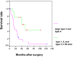
Figure 2
Kaplan–Meier analysis of patients with types 1, 2, 3 (< 80 mm)
and large type-3 and type-4 groups. There was a significant difference in OS
between the two groups (P= 0.0034).
Table 5
Table 5
Clinicopathological features of long-term survivors with CY1 and no other stage IV factor (n = 12).
Discussion
Although the prognosis of CY1 patients is poor, the optimal treatment for these patients has not been established. In the present study, we investigated the clinical outcomes and prognostic factors of 39 patients with CY1/P0 gastric cancer, and identified INF and postoperative chemotherapy as independent prognostic factors for OS. Moreover, INF was found to be an independent prognostic factor for PFS. The percentage of long-term survivors with large type-3 and type-4 tumors was significantly lower compared with those with types 1–3 (< 80 mm). The therapeutic strategy was thought to be very important for CY1/P0 gastric cancer patients with an infiltrative growth pattern such as large type-3 and type-4 tumors to improve prognosis.
According to the Japanese Classification of Gastric Carcinoma, CY should be performed immediately after laparotomy in all patients with gastric carcinoma, except in those with a T1 tumor [6]. Available evidence [9,10,12] suggests that oncologists consider the prognosis of CY1 to be dismal. Fukagawa et al. [12] reported that the 5-year OS rate among 88 patients with CY1/P0 gastric cancer is 7.8%, and five patients experienced recurrence-free survival. Mezhir [9] reported a 5-year OS rate < 10% for patients with M1 Cyt+ (corresponding to CY1/P0 here). In the present study, the 5-year OS rate of the 39 CY1/P0 cases was 10%. Further, postoperative chemotherapy was an independent prognostic factor of OS. Yamashita et al. [14] reported that long-term postoperative adjuvant therapy contributed to a 5-year OS rate of >20% in 35 patients with CY1 and no other stage IV factors (P0H0CY1). Mezhir et al. [9] found that macroscopic peritoneal dissemination is associated with a dismal prognosis (e.g. no survivors >5 years), indicating that the clinical significance of surgical resection of the primary tumor is not established. CY1 is defined as M1 in the latest Japanese treatment guidelines for gastric cancer, which recommends palliative surgery as an option [6].
Mezhir et al. [9] reported a median disease-specific survival for P1 of 0.8 years, and 1.3 years for CY1/P0. Kanazawa et al. [15] demonstrated that the MST by group was 34.5 months for CY1/P0, 34.3 months for CY0/P1, and 19.3 months for CY1/P1, with the OS in CY1/P0 and CY0/P1 groups being significantly longer than in the CY1/ P1 group. These studies indicate that the prognosis of patients with CY1/P0 gastric cancer is better than that of patients with P1 gastric cancer [9 and 15]. Our patients with CY1/P0 gastric cancer included two (5.1%) who survived >5 years with no evidence of recurrence, and one patient who has survived for 10 years and 3 months. Fukagawa et al. [12] reported recurrence-free patients with CY1 advanced gastric cancer who underwent gastrectomy, and showed that five (5.7%) of the 88 patients survived for more than 5 years without evidence of recurrence, indicating that surgery might serve as a promising strategy. In our hospital, surgery was selected as the first-line therapy of patients with CY1/P0 gastric cancer, except four patients with type- 4 disease who underwent neoadjuvant chemotherapy using S-1 and CDDP. However, only one patient survived for more than 3 years.
The optimal treatment strategy for CY1/P0 gastric cancer is controversial. Available evidence suggests that the findings of peritoneal lavage cytology are a strong prognosticator. For example, the median survival times of patients with positive cytology range between 10.5–14.8 months after gastrectomy [16 and 17]. Okabe et al. [18] reported that neoadjuvant chemotherapy is effective for patients with CY1 and an MST of 43.2 months following neoadjuvant therapy. Kodera et al. [19] found that adjuvant chemotherapy with S-1 is effective (MST of 23.5 months). In our present study, the MST of patients with CY1/P0 disease who underwent postoperative chemotherapy using S-1 was 27.0 months. We believe therefore that gastrectomy with adjuvant S-1 might improve survival. Most of our long-term survivors were administered S-1 postoperatively, and 29 patients received postoperative S-1 or S-1 + another drug. Further, of the 12 long-term survivors, after surgery, three patients were administered S-1 only, and six patients were administered S-1 + PTX or + DOC. Adjuvant monotherapy with S-1 or taxane is acceptable and efficient for preventing peritoneal recurrence. In our experience, nine (75.0%) of 12 long-term survivors were administered S-1 or taxane postoperatively. Kanazawa et al. [15] reported the efficacy of adjuvant chemotherapy with S-1 followed by DOC after R1 resection for CY1/P0 gastric cancer. According to the results of their study, this adjuvant chemotherapy is safe and well tolerated and has the potential to improve OS (MST = 34.5 months). Therefore, based on the evidence cited, we consider S-1 or S-1 + taxane effective adjuvant chemotherapy to be effective for CY1/P0 gastric cancer after surgery.
The development of novel chemotherapeutic regimens for intraperitoneal chemotherapy will be challenging. Peritoneal dissemination is among the most important and difficult therapeutic targets in patients with gastric cancer [20]. Although, current treatment outcomes are disappointing for surgery and systemic therapy, intraperitoneal chemotherapy in combination with intravenous chemotherapy shows promise [21]. The rationale for such treatment is based on the slow absorption of paclitaxel from the peritoneum into the body, which maintains a high concentration in the peritoneum [22 and 23]. Therapy using IP/IV paclitaxel + S-1 to treat patients with gastric cancer with macroscopic peritoneal metastasis achieves an MST of 17.6 months and 1-year survival rate >77.1% [21], which is consistent with the results reported here for patients with CY1/P0 disease.
IP/IV therapy may be feasible for patients with CY1 gastric cancer
and for those with preperitoneal dissemination. An alternate strategy
for CY1 or peritoneal dissemination might increase the response rate
as preoperative chemotherapy. Recently, potent chemotherapy in
the form of DCS was administered to patients with advanced gastric
cancers, and achieved the conversion of CY1 to CY0 [24]. Salvage
gastrectomy may serve as an option for patients with CY1 gastric
cancer. For example, such salvage surgery after IP/IV paclitaxel + S1
therapy improved survival compared with standard chemotherapy
such as S1/Cisplatin [25]. The MST of patients with severe peritoneal
metastasis and as cites receiving salvage surgery after IP/IV paclitaxel
+ S1 therapy was 26.4 months [25]. This compares with17.3 months
for patients with large type-3 and type-4 cancer receiving S-1 + CDDP
followed by gastrectomy [26].
In the present study, we treated four patients with type-4 disease
who were administered S-1 + CDDP, and only one survived beyond
3 years. One patient with type-4 disease among the 12 long-term
survivors received neoadjuvant therapy using S-1 + CDDP, and IP/IV
paclitaxel + S-1 + hyperthermia after surgery. One of the three longtime
survivors of large type-3 disease received postoperative DCS
therapy. Recently, Ishigami et al. [27 and 28] reported that intravenous
and intraperitoneal paclitaxel combined with S-1 is very effective
for patients with P1 and/or CY1. In their phase II trial, the MST
and 1-year survival rate was 22.5 months and 78%, respectively, and
peritoneal cytology results turned negative in 24 of 28 patients [27].
This treatment strategy is now under investigation in a phase III
randomized control trial of S-1 plus intravenous and intraperitoneal
paclitaxel versus S-1 plus cisplatin for gastric cancer with peritoneal
metastasis (PHOENIX-GC trial).
Kano et al. [29] found that macroscopic type and residual tumor
status were independent prognostic factors for patients with gastric
cancer. After combining these two factors, no significant difference in
survival was observed between CY1 patients with type-4 tumors who
underwent R1 or R2 resection. In contrast, there was a significant
difference in survival between CY1 patients without type-4 tumors
who underwent R1 or R2 resection [29]. In our present study, the
number of long-term survivors with type 4 and large type-3 cancers
was significantly lower compared with that of long-term survivors
with other macroscopic types.
INFc is an independent predictor of poor prognosis. However,
INF is determined by pathologic examination of the resected stomach
after surgery, and it is therefore difficult to accurately determine INF
status before surgery. In the current study, the number of patients
with large type-3 and type-4 tumors with INFc was 22 (84.6%).
Conversely, there were only three patients with INFc among those
with types 1, 2, and small type-3 disease (23.1%). Univariate analysis
showed that large type-3 and type-4 gastric cancers were associated
with high recurrence rates and poor prognosis for OS and PFS.
However, multivariate analysis did not indicate that macroscopic
type was an independent prognostic factor of disease recurrence.
Kano et al. [29] reported that gastrectomy should be avoided for
treating patients with type-4 tumors with overt peritoneal metastasis
or with positive peritoneal cytology. In Kano’s previous series, 22
patients with type-4 tumors underwent staging laparoscopy, and
10 (45.4%) were diagnosed with overt or occult peritoneal disease
[29]. The information provided by CY is therefore essential for
determining the therapeutic strategy for patients with type-4 gastric
cancer. Therefore, we recommend that staging laparoscopy should
be included among the first procedures performed. In our present
study, the prognosis of patients with CY1 with large type-3 and
type-4 tumors was extremely poor, even when an R1 resection was
performed. Miki et al. [30] reported that staging laparoscopy reveals
peritoneal metastasis in 36.3% of patients with large type-3 and
type-4 gastric cancers without clinically evident factors that indicate
incurable disease such as malignant ascites and/or distant metastasis.
Patients with type-4 or large (tumor diameter ≥8 cm) type-3 gastric
cancer are considered suitable candidates for staging laparoscopy
because of the high incidence of peritoneal metastasis. Staging
laparoscopy is considered to be a useful tool in detecting unsuspected
peritoneal metastasis not observed by other diagnostic imaging such
as abdominal ultrasonography or abdominal computed tomography
[29]. Patients with cM0 type-4 and large type-3 gastric cancers are
considered suitable candidates for staging laparoscopy [29]. Because
the prognosis of patients with large type-3 and type-4 cancers with
CY1/P0 is very poor, they likely require staging laparoscopy. Further,
neoadjuvant chemotherapy such as DCS or IP/IV paclitaxel should be
considered for treating patients with CY1 gastric cancer even without
overt peritoneal dissemination.
The present study has some limitations. First, this was a
retrospective investigation with patients treated at a single institution
during a 15-year period (2000–2014), and the diagnostic modalities,
operative procedures, and pre- and postoperative adjuvant therapy
varied. Nevertheless, our study provides compelling evidence
indicating that patients with large type-3 and type-4 CY1/P0 gastric
cancer require multidisciplinary therapy such as neoadjuvant
chemotherapy, using DCS and IP/IV paclitaxel.
Conclusion
INF and postoperative chemotherapy are independent prognostic factors for OS in gastric cancer patients. Moreover, INF is also an independent prognostic factor for PFS. However, INF is only determined postoperatively by pathological examination of the resected stomach. Macroscopic type is not an independent prognostic or recurrence factor. Univariate analysis showed that large type 3 and type 4 cancers are associated with a high recurrence rate and poor prognosis in terms of both OS and PFS. Moreover, the number of large type 3 and type 4 cases with INFc was very high. Therefore, these cases are considered to require multidisciplinary therapy, including neoadjuvant chemotherapy.
References
- International Agency for Research on Cancer WHO. GLOBOCAN 2012: Estimated cancer incidence, mortality and prevalence 2012.
- Sakuramoto S, Sasako M, Yamaguchi T, Kinoshita T, Fujii M, Nashimoto A. Adjuvant chemotherapy for gastric cancer with S-1, an oral fluoropyrimidine. N Engl J Med. 2007; 357: 1810–1820.
- Yamashita K, Sakuramoto S, Kikuchi S, Katada N, Kobayashi N, Watanabe M. Validation of staging systems for gastric cancer. Gastric Cancer. 2008; 11: 111–118.
- Bringand C, Arviex C, Gilly FN, Glehen O. Treatment of peritoneal carcinomatosis in gastric cancers. Dig Dis. 2004; 22: 366–373.
- Isobe Y, Nashimoto A, Akazawa K, Oda I, Hayashi K, Miyashiro I, et al. Gastric cancer treatment in Japan: 2008 annual report of the JGCA nationwide registry. Gastric Cancer. 2011; 14: 301–316.
- Japanese Gastric Cancer Association. Japanese classification of gastric carcinoma: 3rd English edn. Gastric Cancer. 2011; 14: 101–112.
- Sobin L, Gospodarowicz M, Wittekind C. TNM classification of malignant tumours. 7th edn. New York; Wiley-Blackwell. 2009.
- Leake PA, Cardoso R, Seevaratnam R, Lourenco L, Helyer L, Mahar A, et al. A systematic review of the accuracy and utility of peritoneal cytology in patients with gastric cancer. Gastric Cancer. 2012; 15: 27–32.
- Mezhir JJ, Shah MA, Jacks LM, Brennan MF, Coit DG, Strong VE. Positive peritoneal cytology in patients with gastric cancer: natural history and outcome of 291 patients. Ann SurgOncol. 2010; 17: 3173–3180.
- Lee SD, Ryu KW, Eom BW, Lee JH, Kook MC, Kim YW. Prognostic significance of peritoneal washing cytology in patients with gastric cancer. Br J Surg. 2012; 99: 397–403.
- Japanese Gastric Cancer Association. Japanese classification of gastric carcinoma: 2nd English Edition. Gastric Cancer. 1998; 1: 10–24.
- Fukagawa T, Katai H, Saka M, Morita S, Sasajima Y, Taniguchi H, et al. Significance of lavage cytology in advanced gastric cancer patients. World J Surg. 2010; 34: 563–568.
- Japanese Gastric Cancer Association. Japanese gastric cancer treatment guidelines?. 2010 (ver. 3). Gastric Cancer. 2011; 14: 113–123.
- Yamashita K, Ushiku H, Katada N, Hosoda K, Moriya H, Mieno H, et al. Reduced preoperative serum albumin and absence of peritoneal dissemination may be predictive factors for long-term survival with advanced gastric cancer with positive cytology test. Eur J SurgOncol. 2013; 39: 1309–1316.
- Kanazawa Y, Kato S, Fujita I, Onodera H, Uchida E. Adjuvant chemotherapy with S-1 followed by docetaxel for gastric cancer and CY1P0 peritoneal metastasis after relatively curative surgery. J Nippon Med Sch. 2013; 80: 378–383.
- Cabalag CS, Chan ST, Kaneko Y, Duong CP. A systematic review and metaanalysis of gastric cancer treatment in patients with positive peritoneal cytology. Gastric Cancer. 2015; 18: 11–22.
- De Andrade JP, Mezhir JJ. The critical role of peritoneal cytology in staging of gastric cancer: an evidence-based review. J Surg Oncol. 2014; 110: 291– 297.
- Okabe H, Ueda S, Obama K, Hosogi H, Sakai Y. Induction chemotherapy with S-1 plus cisplatin followed by surgery for treatment of gastric cancer with peritoneal dissemination. Ann SurgOncol. 2009; 16: 3227–3236.
- Kodera Y, Ito S, Mochizuki Y, Kondo K, Koshikawa K, Suzuki N, et al. A phase II study of radical surgery followed by postoperative chemotherapy with S-1 for gastric carcinoma with free cancer cells in the peritoneal cavity (CCOG0301 study). Eur J Surg Oncol. 2009; 35: 1158–1163.
- Yamashita K, Sakuramoto S, Kikuchi S, Katada N, Kobayashi N, Watanabe M. Surgical resection of stage IV gastric cancer and prognosis. Anticancer Res. 2007; 27: 4381–4386.
- Yamaguchi H, Kitayama J, Ishigami H, Emoto S, Yamashita H, Watanabe T. A phase 2 trial of intravenous and intraperitoneal paclitaxel combined with S-1 for treatment of gastric cancer with macroscopic peritoneal metastasis. Cancer. 2013; 119: 3354–3358.
- Francis P, Rowinsky E, Schneider J, Hakes T, Hoskins W, Markman M. Phase I feasibility and pharmacologic study of weekly intraperitoneal paclitaxel: a Gynecologic Oncology Group pilot study. J Clin Oncol. 1995; 13: 2961–2967.
- Armstrong DK, Bundy B, Wenzel L, Huang HQ, Baergen R, Lele S, et al. Intraperitoneal cisplatin and paclitaxel in ovarian cancer. N Engl J Med. 2006; 354: 34–43.
- Koizumi W, Nakayama N, Tanabe S, Sasaki T, Higuchi K, Nishimura K. A multicenter phase II study of combined chemotherapy with docetaxel, cisplatin, and S-1 in patients with unresectable or recurrent gastric cancer (KDOG0601). Cancer Chemother Pharmacol. 2012; 69: 407–413.
- Kitayama J, Ishigami H, Yamaguchi H, Yamashita H, Emoto S, Kaisaki S, et al. Salvage gastrectomy after intravenous and intraperitoneal paclitaxel (PTX) administration with oral S-1 for peritoneal dissemination of advanced gastric cancer with malignant ascites. Ann Surg Oncol. 2014; 21: 539–546.
- Iwasaki Y, Sasako M, Yamamoto S, Nakamura K, Sano T, Katai H, et al. Phase II study of preoperative chemotherapy with S-1 and cisplatin followed by gastrectomy for clinically resectable type 4 and large type 3 gastric cancer (JCOG0210). J SurgOncol. 2013; 107: 741–745.
- Ishigami H, Kitayama J, Kaisaki S, Hidemura M, Kato K, Otani T, et al. Phase II study of weekly intravenous and intraperitoneal paclitaxel combined with S-1 for advanced gastric cancer with peritoneal metastasis. Ann Oncol. 2010; 21: 67–70.
- Ishigami H, Kitayama J, Otani K, Kamei T, Soma D, Miyato H, et al. Phase I pharmacokinetic study of weekly intravenous and intraperitoneal paclitaxel combined with S-1 for advanced gastric cancer. Oncology. 2009; 76: 311–314.
- Kano Y, Kosugi S, Ishikawa T, Otani T, Muneoka Y, Sato Y, et al. Prognostic significance of peritoneal lavage cytology at three cavities in patients with gastric cancer. Surgery. 2015; 158: 1581–1589.
- Miki Y, Tokunaga M, Tanizawa Y, Bando E, Kawamura T, Terashima M. Staging laparoscopy for patients with cM0, type 4, and large type 3 gastric cancer. World J Surg. 2015; 39: 2742–2747.