Case Report
Left Ventricular Mass: Myxoma or Thrombus: Resection Through the Mitral Valve via the Transeptal Approach
Artemiou P1*, Martincek M2, Gasparovic I1 and Schusterova I3
1Department of Cardiac Surgery, Slovak Medical University, Slovakia
2Department of Cardiac Surgery, University Jan Pavel Safarik, Slovakia
3Department of Cardiac Surgery, Children’s Faculty Hospital, Slovakia
*Corresponding author: Panagiotis Artemiou, Department of Cardiac Surgery, National Institute for Cardiovascular Diseases, Slovak Medical University, Pod Krasnou Horkou 1, 83101 Bratislava, Slovakia
Published: 31 Aug, 2016
Cite this article as: Artemiou P, Martincek M, Gasparovic I, Schusterova I. Left Ventricular Mass: Myxoma or Thrombus: Resection Through the Mitral Valve via the Transeptal Approach. Clin Surg. 2016; 1: 1094.
Abstract
In this case report a 43 years old male patient was presented with symptoms of embolic right leg
ischemia. Transthoracic echocardiography showed a left ventricular mass present toward the apex
attached to the interventricular septum with the presumptive diagnosis of myxoma. He underwent
a successful removal of the tumor through the mitral valve via the transeptal approach. Histology
showed mixed thrombus.
Keywords: Left ventricular mass; Transeptal approach; Myxoma; Thrombus
Introduction
Patient with embolic episode should always be evaluated for cardiac masses. Mass in the Left Ventricle (LV) can be a myxoma or a thrombus. In either case, mobile mass with embolic potential should be surgically resected [1-4]. Here we report a case of a left ventricular thrombus that was supposed to be myxoma preoperatively undergoing successful resection presenting as an embolic lower limb ischemia.
Case Presentation
A 43 years old smoker male patient with a negative medical history was admitted due to calf
pain and numbness of the toes of the right leg for the last ten days. Laboratory, chest X-ray and ECG
examinations were normal. Clinical examination of the right leg showed absent peripheral pulses of
the dorsal pedis and tibial posterior arteries.
During hospitalization, transthoracic echocardiographic (TTE) examination showed a 1.6 x 1.7
(Figure 1) pediculated hyperechogenic hypermobile mass present in the Left Ventricle (LV) toward
apex attached to the interventricular septum. No LV regional wall motion abnormality was detected
with LV ejection fraction of 60%. The mass was presumed to be myxoma and considering the clinical
presentation of the patient and the potential for further embolization the patient was scheduled for
an emergency open heart surgery. For the leg embolism conservative treatment was recommended.
A median sternotomy was performed. The ascending aorta and both venae cavae were
cannulated and standard cardiopulmonary bypass (CPB) was performed. The heart was stopped by
cross-clamping the ascending aorta. Myocardial protection was achieved by means of intermittent
anterograde administration of cold blood cardioplegia. The venae cavae were snared with tourniquets
and via the transeptal approach through the mitral valve a fragile jelly-like mass was visible attached
to the interventricular septum towards the apex. With care not to injure the valve, chordae tendinae
and papillary muscle the mass was completely removed through the mitral valve and the tumor’s
pedicle was completely shaved from the endocardium (Figure 2). The patient was weaned from
CPB easily, without inotropic support. The postoperative course was uneventful, the patient was
put on anticoagulation treatment and on the postoperative day 7 he was discharged home. The
postoperative TTE was normal. Histopathologic examination showed a mixed thrombus, partially
organized (Figure 3). An informed consent was obtained from the patient to present the case.
Discussion
Benign cardiac myxomas constitute of 88% of cardiac tumor cases and LV myxomas account for
only 2.5% of cases [4]. The first reported case of surgical removal of a LV myxoma was published by
Kay et al. [5] in 1959. Since, then at least 71 other surgical cases of LV myxoma usually originated
from the interventricular septum have been reported mostly as single
cases [6].
The clinical features are mostly caused by embolization and
obstruction of the left ventricular outflow tract [7]. Embolic
phenomena are more common than LA myxoma, occurring in 64% of
patients with LV myxoma [8]. Presenting symptoms of embolic lower
limb ischemia are also reported by other authors [9]. Considering the
risk of embolization, myxomas should be surgically resected as early
as possible.
Thrombus formation in LV is a known complication of heart
failure and acute myocardial infarction [10,11]. Hyrecoagulable
state or undetectable disorder of the endocardium can lead to
ventricular thrombus formation even in normal heart [12]. Main
causes of inherited thrombophilia such as, factor V gene mutation,
prothrombin gene mutation, antithombin and protein C and S are
also associated with LV thrombus formation [13]. Left ventricular
thrombus formation is also associated with the antiphospholipid
antibody syndrome [14], the autoimmune disorders of Behcet disease
and lupus erythematosus [15,16].
The differential diagnosis of a LV mass between thrombus and
myxoma can be challenging. However, there is no diagnostic feature,
either by 2D-echocardiography or by direct inspection in which the
diagnosis can be confirmed, and either pathology may masquerade
the other [17]. Histopathology is the final court of appeal and must
always be performed, like in our report.
The surgical approach we chose to remove the tumor in this
location was though the right atrium and atrial septum, but can be
carried also by other ways. 1) Through the left atrium and mitral valve
[18]. 2) Through the ascending aorta with video assistance [19,20]. 3)
Through a small longitudinal incision in the left ventricle [6,21].
We have chosen the transeptal approach because a) Avoids
ventriculotomy and its potential complications like deterioration of
the LV function and bleeding, despite the fact that ventriculotomy
provides good visibility and the possibility for complete resection.
b) Through this approach exploration of the right, left atrium and
interatrial septum if necessary can be done.
A potential disadvantage of the approach through the mitral valve
would be limited room for maneuvering during the mass resection.
Recently, endoscopy and minimally invasive techniques have also
been applied [22,23].
First line treatment for a LV thrombus is anticoagulation,
however a mobile thrombus presented as an embolic complication as
in this case often requires urgent surgical resection.
In conclusion we reported a successful resection of LV thrombus
present toward the apex attached to the interventricular septum with
the presumptive preoperative diagnosis of myxoma through the
mitral valve via the transeptal approach. The patient was discharged
home in good condition.
Figure 1
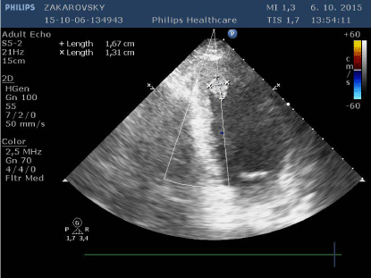
Figure 1
Transthoracic echocardiogram reveals the tumor in the left
ventricular cavity attached to the interventricular septum.
Figure 2
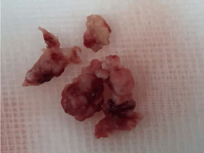
Figure 2
Photograph shows the resected mass.
Figure 3
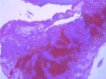
Figure 3
Histologic examination: Mixed thrombus, partially organized,
Hematoxylin and eosin stain.
References
- Sarjeant JM, Buttany J, Cusimano RJ. Cancer of the heart: Epidemiology and management of primary tumors and metastases. J Am Cardiovasc Drugs. 2003; 3: 407-421.
- Meller J, Teicholz LE, Pichard AD. Combination of right atrial mass and left ventricular myxoma: Echocardiographic diagnosis and review of the literature. Am J Med. 1977; 63: 816-823.
- Kumar P, Garg A. Left ventricular myxoma in a child: a case report. Eur J Echocardiogr. 2011: 12: E23.
- Korkmaz AA, Tamtekin B, Onan B, Demir AS, Guden M, Uckurt Y, et al. Combination of left atrial and left ventricular myxoma. Ann Thorac Surg. 2010; 89: 33-35.
- Kay JH, Anderson RM, Meihaus J, Lewis R, Magidson O, Bernstein S, et al. Surgical removal of an intracavitary left ventricular myxoma. Cirulation. 1959; 20: 881-886.
- Abad C, Novoa J, Delgado A, Alonso A. Myxoma of the left ventricle. Tex Heart Inst J. 2014; 41: 395-400.
- Mobeirek AF, Al-Nozha M. Multiple left ventricular myxoma. Case report and review of the literature. J Saudi Hean Assoc. 1996; 8: 122-126.
- Samdarshi TE, Mahan EF, Nanda SC, Guthrie FW, Bernstein IJ, Kiklin JW. Transesophageal echocardiography diagnosis of multicentric left ventricular myxomas mimicking a left atrial tumor. J Thorac Cardiovasc Surg. 1992; 103: 471-474.
- Prifti E, Adernaj F, Kajo E, Babosi A. A giant myxoma originating from the aortic valve causing severe left ventricular tract obstruction: a case report and literature review. World J Surg Oncol. 2015; 13: 151.
- Kalra A, Jang IK. Prevalence of left ventricular thrombus after primary coronary intervention for acute myocardial infarction. J Thromb Thrombolysis. 2000; 10: 133-136.
- Gottdiener JS, Gay JA, Van-Voorhees L, Di-Bianco R, Fletcher RD. Frequency and embolic potential of left ventricular thrombus in dilated cardiomyopathy. Assessment by 2-dimensional echocardiography. Am J Cardiol. 1983; 52: 1281-1285.
- Verma AK, Alam M, Rosman HS, Brymer J, Keith F. Systemic embolization from thrombus in normal left ventricles. Chest. 1988; 93: 441-442.
- Lane DA, Mannucci PM, Bauer HS, Bertina RM, Bochkov NP, Boulyjenkov V, et al. Inherited thrombophilia: Part I. Thromb Haemost. 1996; 76: 651-662.
- Aguilar JA, Summerson C. Intracardiac thrombus in antiphospholipid antibody syndrome. J Am Soc Echocardiogr. 2000; 13: 873-875.
- Vanhaleweyk G, El-Ramahi KM, Hazmi M, Sieck JO, Zaman L, Fawzy M. Right atrial, right ventricular and left ventricular thrombi in (incomplete) Behcet’s disease. J. Oxford. 1990; 11: 957-959.
- Barjatiya MK, Shah NK, Kothan SS, Shah PP, Trivedi HL. Spontaneous left ventricle cavity thrombus in a patient of systemic lupus erythematosus. J Assoc Physicians India. 1992; 40: 195-196.
- Kazmierczak M, Kazmierczak E, Sarnowska Z. Left ventricular thrombus imitating cardiac myxoma, successfully treated with clopidogrel. A case report. Kardiol Pol. 2001; 55: 333-335.
- Tanaka D, Unai S, Diehl JT, Hiroshe H. Surgical removal of a large mobile left ventricular thrombus via left atriotomy. World J Clin Cases. 2014; 2: 32-35.
- Qin W, Wang L, Chen X, Liu P, Wang R. Left ventricular myxoma: a case report. J Biomed Res. 2014; 28: 506-508.
- Tsukube T, Okada M, Ootaki Y, Tsuji Y, Yamashita C. Trans-aortic videoassisted removal of a left ventricular thrombus. Ann Thorac Surg. 1999; 68: 1063-1065.
- Raut MS, Maheshwari A, Dubey S, Joshi S. Left ventricular mass: Myxoma or thrombus? Ann Card Anaesth. 2015; 18: 95-97.
- Hasan M, Smith JM. Robotic assisted excision of a left ventricular myxoma. Interact Cardiovasc Thorac Surg. 2012; 14: 113-114.
- Kammerer I, Nagib R, Franke UFW. Minimal invasive thoracoscopic transmitral resection of a left ventricular myxoma. Arch Clin Exp Surg. 2012; 42: 502-504.