Case Report
Mucosa Associated Lymphoid Tissue Lymphoma (Maltoma) of Parotid Gland
Dokuzlar U* Mıman MC, Egrilmez M and Denızoglu II
Department of Otolaryngology, Izmir University, Turkey
*Corresponding author: Ugur Dokuzlar, Department of Otolaryngology, Izmir University, Yeni Girne Blv. 1825 sk. No: 12, Karsiyaka, Izmır, Turkey
Published: 29 Aug, 2016
Cite this article as: Dokuzlar U, Mıman MC, Egrilmez M, Denızoglu II. Mucosa Associated Lymphoid Tissue Lymphoma (Maltoma) of Parotid Gland. Clin Surg. 2016; 1: 1089.
Abstract
Mucosa-Associated Lymphoid Tissue (MALT) is a no encapsulated cluster of lymphocytes that is
thought to have important role in mucosal immunity. Maltoma is an extranodal lymphoma either
of B- or T-cell origin that may most common involve stomach, followed by other gastrointestinal
organs, respiratory tract, skin and others containing lymphoid tissue parts. Maltoma in salivary
gland is a rare entity because it is not a part of salivary gland tissue, but chronic inflammatory
processes can cause lymphocyte accumulation in gland and these cells can be cause of Maltomas.
Tumors located in parotid gland are reported to be malignant in 26-32% of cases. Physical
examination does not effective to rule out malignancy in all cases. Preoperative studies including
imaging modalities such as ultrasonography (US) and Magnetic Resonance Imaging (MRI), and
histopathological studies (Fine Needle Aspiration Cytology (FNAC)) are used to have accurate
preoperative diagnosis.
In this case, we report a rare tumor of parotid gland in a patient with breast cancer history that is
confusing clinical situation and the insufficiency of preoperative and peroperative diagnostic studies
to determine the pathology in parotid gland.
Despite improvement of diagnostic studies personal experience and evaluation of clinician remains
most important part of planning of treatment strategies for parotid gland neoplasm.
Introduction
Mucosa-Associated Lymphoid Tissue (MALT) is a no encapsulated cluster of lymphocytes
that is thought to have important role in mucosal immunity [1]. In 1983 Isaacson and Wright
described lymphoma arising from MALT (Maltoma) and it was recognized as a discrete entity in
1994 by the Revised European-American Lymphoma classification [2]. Maltoma is an extranodal
lymphoma either of B- or T-cell origin that may most common involve stomach, followed by other
gastrointestinal organs, respiratory tract, skin and others containing lymphoid tissue parts [3-7].
Any local chronic inflammatory process (autoimmune diseases, infections) may induce lymphoid
proliferation and suspected to be in pathogenesis of Maltoma [8,9]. In general Maltoma is a low
grade, localized tumor and usually does not metastasize at early periods [10].
Maltoma in salivary gland is a rare entity because lymphoid tissue is not a part of salivary gland,
but chronic inflammatory processes like Sjogren’s Disease and Hashimoto’s Thyroiditis can cause
lymphocyte accumulation in gland and these cells can be cause of Maltomas [8,10,11]. In Sjogren’s
Disease (SD) periductal lymphoepithelial islands consist of reactive, polyclonal cells are seen in
parotid gland and normal lobular appearance of the gland is preserved [11]. There is an increased
risk of malignant lymphomas in chronic autoimmune disorders and in SD this risk is approximately
44 times higher than normal population [12]. It is seen in biochemistrical analysis of gland biopsies
that immunoglobulin M bearing plasma cells are increased in glands and this suggests possibility
of monoclonal expansion of lymphocytes in SD. If monotypic plasma cell is found in gland tissue
patient has higher risk of lymphoproliferative disorders [13].
Tumors located in parotid gland are reported to be malignant in 26-32% of cases [14]. Physical
examination does not effective to rule out malignancy in all cases. Preoperative studies including
imaging modalities such as ultrasonography (US) and Magnetic Resonance Imaging (MRI), and
histopathological studies (Fine Needle Aspiration Cytology (FNAC)) are used to have accurate
preoperative diagnosis [14].
Metastasis to parotid gland is a common reason for parotid gland masses. In a recent study it
was reported that in 89 patients with malignant tumors of parotid
gland, 39 (44%) were metastatic [15]. Although majority of primary
tumor in this these cases located in head and neck region, metastasis
of renal cell carcinoma, breast carcinoma and carcinoma of bronchus
was reported in this study.
In this case, we report a rare tumor of parotid gland in a patient
with breast cancer history that is confusing clinical situation and the
insufficiency of preoperative and peroperative diagnostic studies to
determine the pathology in parotid gland.
Figure 1
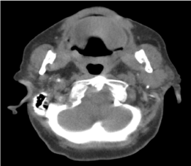
Figure 1
CT image of parotid mass extending to the deep lobe.
Case Presentation
A 69-year-old woman presented with a growing mass over left
parotid gland for about a month. Patient had a breast cancer and
right partial mastectomy history, 10 years ago. After the operation
she had been treated by adjuvant radiotherapy and chemotherapy.
Six years after the first treatment a local recurrence observed during
routine follow ups and right radical mastectomy procedure had
been added. Adjuvant chemotherapy (trastuzumab) had been given
after surgery for one year. Patient still uses letrozole since her last
operation. A Computed Tomography (CT) scanning had been done
in another medical center before she referred to our clinic. CT
showed a lobulated, hyperdense parotid gland mass, containing
microcalcification areas and extending to deep lobe (Figure 1). US
and MRI scanning and FNAB offered to patient, but she refused MRI.
US showed bilateral atrophy in submandibular gland, and a solitary,
hypoechoic 30 mm in diameter mass with hyperechoic foci in left
parotid gland. FNAB reported as Warthin’s tumor. After these results
surgical procedure was planned.
In operation main truncus of facial nerve was found. While
dissecting superficial lobe of gland it was seen that buccal branch of
facial nerve was wrapped by tumor and tumor was going through the
deep lobe. The buccal branch was sacrificed and remaining branches
of facial nerve dissected carefully and superficial lobe taken out.
Incisional sample taken from deep lobe tumor and send for frozen
section evaluation together with superficial lobe. After total dissection
of facial nerve over the tumor, deep lobe of the parotid was removed.
A frozen section result showed no malignancy and operation was
concluded. She had grade IV (House Brackmann Scale) facial nerve
palsy developed in early postoperative period and after 6 months
facial palsy completely recovered.
Pathology department reported “extranodal marginal zone
lymphoma (maltoma)”, with immunohistochemical stains finding
the lymphoid cells to be positive for CD20, bcl-2 after paraffin block
evaluation. The Ki67 was estimated at only 7% of the lesion. Tumor
free areas of gland showed changes suggesting SD (Figure 2).
After pathological results patient reexamined for symptoms of SD
and we found that the patient had complaint about dryness of eyes for
one year and this symptom had been attributed to medication history
for breast cancer by her oncologist.
The patient was informed about her pathological result and we
offered her chemotherapy for maltoma. She refused chemotherapy
because of her treatments for breast cancer. She comes for routine
follow up physical examinations and as a radiological examination
she accepts US alone and she has not any problem in 14th month of
postoperative period.
Figure 2
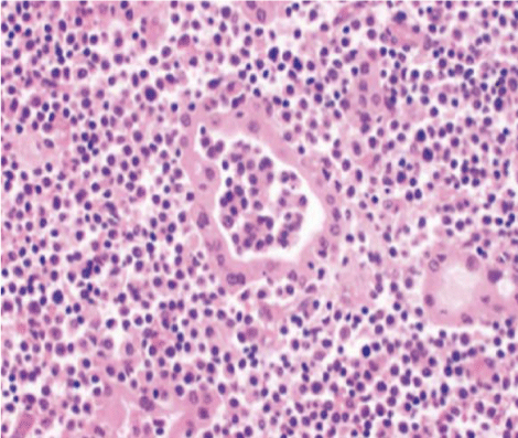
Figure 2
Microscopic appereance of tumor.
Discussion
Because of the slow-growing nature of maltoma expected survival
rates can be observed even in advanced cases [16]. This survival
rates mandates surgeon to select lower morbidity procedures in this
case. Although we need pathological proof of histology of the mass
before the decision of treatment modality, this may not be available
in some cases for parotid gland. Insicional biopsy is contraindicated
for parotid gland masses because of the risk of tumor insemination
and facial nerve injury [17]. FNAC is an evaluation study that can
be easily done with almost no pain to determine whether the mass is
benign or malignant preoperatively. In the literature the sensitivity
and specificity of FNAC is reported in range of 54-92% and 86-100%
respectively [18-20]. Cytological analysis of hypercellular tumors
is particularly difficult. Frozen Section (FS) is a study, performed
peroperative and reported sensitivity and specificity are between 77-
93% and 95-100% respectively [14].
Like as other soft tissue masses, MRI is the imaging method of
choice for the evaluation of parotid tumors. In the recent literature,
the sensitivity of parotid FNAC in the diagnosis of malignancy has
been reported to be in the range of 54–92%, with specificity in the
range of 86–100% [18,19]. The false-negative rate ranges from 2% to
31% and the false-positive rate from 0% to 7% [14]. In our case we
could not have MRI because of patient’s refusal.
FS is a good diagnostic tool to have information on the nature of
the tumor whether it is malignant or not during surgery [14]. Despite
it is a very effective method in most body parts its accuracy in salivary
gland masses is controversial. Therapeutic decision should not be
based on FS solely. Therefore surgeon must consider frozen section
in conjunction with the preoperative clinical findings, diagnostic tests
and patient’s medical history.
Like as other tissues intraparotid metastases can occur many
years after primary tumor treatment, and can be the first clinical
manifestation of a recurrence. Parotidectomy with complete excision
of the tumor can be curative [15] and is necessary for exact diagnosis
in most cases. In our case we could have pathological diagnosis after
total excision of gland.
In this case we reported fail of both preoperative and peroperative
diagnostic studies in a patient with a confusing previous malignancy
history. In our case we decided to perform total parotidectomy
because of patient’s breast cancer history and infiltrating feature of
the mass made us think that it could be malignant. Perineurium is a
well-known barrier to tumor invasion, and this allowed us to protect
facial nerve because of we had no certain diagnosis of malignancy. FS
result confirmed our decision and we protected patient’s facial nerve
branches those were not affected. In fact our patient refused further
therapies for Maltoma and we follow up her by physical examination
and US alone according to her request.
Conclusions
Despite improvement of diagnostic studies personal experience and evaluation of clinician remains most important part of planning of treatment strategies for parotid gland neoplasm.
References
- Ciccone E, Truini M, Grossi CE. Lymphoid complement of the human salivary glands: Function and pathology. Eur J Morphol. 1998; 36: 252-256.
- Isaacson P, Wright DH. Malignant lymphoma of mucosa associated lymphoid tissue: A distinctive type of B-cell lymphoma. Cancer. 1983; 52: 1410-1416.
- Thieblemont C, Berger F, Dumontet C, Moullet I, Bouafia F, Felman P, et al. Mucosa-associated lymphoid tissue lymphoma is a disseminated in one third of 158 patients analyzed. Blood. 2000; 95: 802-806.
- Zucca E, Conconi A, Pedrinis E, Cortelazzo S, Motta T, Gospodarowicz MK, et al. Non-gastric marginal zone B-cell lymphoma of the mucosa associated lymphoid tissue. Blood. 2003; 101: 2489-2495.
- Zinzani PL, Magagnoli M, Galieni P, Martelli M, Poletti V, Zaja F, et al. Nongastrointestinal low grade mucosa-associated lymphoid tissue lymphoma: Analysis of 75 patients. J Clin Oncol. 1999; 117: 1254-1258.
- Tsang RW, Gospodarowicz MK, Pintilie M, Wells W, Hodgson DC, Sun A, et al. Localized mucosa associated lymphoid tissue lymphoma treated with radiation therapy has excellent clinical outcome. J Clin Oncol. 2003; 21: 4157-4164.
- Wickramasinghe A, Howarth A, Drage NA. Multiple bilateral parotid sialoliths in a patient with mucosa-associated lymphoid tissue lymphoma (MALT lymphoma) of the salivary glands. Oral Surg Oral Med Oral Pathol Oral Radiol Endod. 2005; 99: 496-498.
- Thieblemont C, Bastion Y, Berger F, Rieux C, Salles G, Dumontet C, et al. Mucosa-associated lymphoid tissue gastrointestinal and nongastrointestinal lymphoma behavior: Analysis of 108 patients. J Clin Oncol. 1997; 15: 1624-1630.
- Zucca E, Roggero E, Bertoni F, Conconi A, Cavalli F. Primary extranodal non-Hodgkin’s lymphomas. Part 2: Head and neck, central nervoussystem and other less common sites. Ann Oncol. 1999; 10: 1023-1033.
- Anacak Y, Miller RC, Constantinou N, Mamusa AM, Epelbaum R, Li Y, et al. Primary mucosa-associated lymphoid tissue lymphoma of the salivary glands: a multicenter Rare Cancer Network study. Int J Radiat Oncol Biol Phys. 2012; 82: 315-320.
- Ando M, Matsuzaki M, Murofushi T. Mucosa-associated lymphoid tissue lymphoma presented as diffuse swelling of the parotid gland. Am J Otolaryngol. 2005; 26: 285-288.
- Kassan SS, Thomas TL, Moutspoulos HM, Hoover R, Kimberly RP, Budman DR, et al. Increased risk of lymphoma in sicca syndrome. Ann Intern Med. 1978; 89: 888-892.
- Bodeutsch C, de Wilde PC, Kater L, van den Hoogen FH, Hene RJ, van Houwelingen JC, et al. Monotypic plasma cells in labial salivary glands of patients with Sjörgren’s syndrome: prognosticator for systemic lymphoproliferative disease. J Clin Pathol. 1993; 46: 123-128.
- Fakhry N, Santini L, Lagier A, Dessi P, Giovanni A. Fine needle aspiration cytology and frozen section in the diagnosis of malignant parotid tumours. Int J Oral Maxillofac Surg. 2014; 43: 802-805.
- Franzen AM, Günzel T, Lieder A. Parotid gland metastases of distant primary tumours: A diagnostic challenge. Auris Nasus Larynx. 2016; 43: 187-191.
- Vazquez A, Khan MN, Sanghvi S, Patel NR, Caputo JL, Baredes S, et al. Extranodal marginal zone lymphoma of mucosa-associated lymphoid tissue of the salivary glands: a population-based study from 1994 to 2009. Head Neck. 2015; 37: 18-22.
- Bussu F, Parrilla C, Rizzo D, Almadori G, Paludetti G, Galli J. Clinical approach and treatment of benign and malignant parotid masses, personal experience. Acta Otorhinolaryngol Ital. 2011; 31: 135-143.
- Fakhry N, Antonini F, Michel J, Penicaud M, Mancini J, Lagier A, et al. Fine-needle aspiration cytology in the management of parotid masses: evaluation of 249 patients. Eur Ann Otorhinolaryngol Head Neck Dis. 2012; 129: 131-135.
- David O, Blaney S, Hearp M. Parotid gland fine-needle aspiration cytology: an approach to differential diagnosis. Diagn Cytopathol. 2007; 35: 47-56.
- Lin AC, Bhattacharyya N. The utility of fine needle aspiration in parotid malignancy. Otolaryngol Head Neck Surg. 2007; 136: 793-798.