Research Article
Intestinal Intraluminal Glycerol and Plasma I-FABP Levels In Preterm Infants with Necrotizing Enterocolitis
Högberg N1 *, Per-Ola Carlsson2,4, Hillered L3, Stenbäck A1, Larsson A4 and Lilja HE1
1Department of Women´s and Children´s Health, Uppsala University, Sweden
2Department of Medical Cell Biology, Uppsala University, Sweden
3Department of Neuroscience, Uppsala University, Sweden
4Department of Medical Sciences, Uppsala University, Sweden
*Corresponding author: Niclas Högberg, Department of Pediatric Surgery, University Children's Hospital, SE-751 85 Uppsala, Sweden
Published: 26 Aug, 2016
Cite this article as: Högberg N, Per-Ola Carlsson, Hillered L, Stenbäck A, Larsson A, Lilja HE. Intestinal Intraluminal Glycerol and Plasma I-FABP Levels In Preterm Infants with Necrotizing Enterocolitis. Clin Surg. 2016; 1: 1085.
Abstract
Background/Purpose: Necrotizing enterocolitis (NEC) is highly associated with prematurity, and is characterized by bowel necrosis and multiple organ failure. There is a strong need for improved
diagnostic methods to reduce the significant morbidity and mortality associated with NEC. The
aim of this single center prospective study was to investigate the possibility of detecting early signs
of NEC, by using rectal intraluminal microdialysis and plasma intestinal fatty acid binding protein
(I-FABP) in preterm infants, admitted to a level III neonatal intensive care unit.
Methods: The study was performed on extremely preterm infants with a gestational age of less than
28 weeks. During a 4-week period after birth, rectal intraluminal microdialysate levels of glucose,
lactate, pyruvate and glycerol were measured, and plasma was collected for I-FABP analysis. Infants
not developing NEC served as controls.
Results: Microdialysis revealed signs of intestinal hypoxic or ischemic damage and cell membrane
degradation, with a marked increase of both intraluminal glycerol and plasma I-FABP in infants
developing NEC, as well as in infants suffering from other complications. The microdialysate levels
of glucose, lactate and pyruvate were too low to be evaluated in this setting. All infants tolerated the
microdialysis well without any complications.
Conclusion: Elevated levels of intraluminal glycerol and plasma I-FABP suggests mucosal cell
membrane degradation and hypoxic or ischemic damage in preterm infants developing NEC, as
well as in preterm infants suffering from other complications such as volvulus, sepsis or respiratory
distress. However, it was not possible to predict development of NEC before clinical diagnosis using
these markers.
Keywords: Necrotizing; Enterocolitis; Intraluminal; Microdialysis; Glycerol; I-FABP
Introduction
Necrotizing enterocolitis (NEC) is the most common gastrointestinal disorder in extremely
premature very low birth weight neonates (VLBW <1500 g) [1]. In this group, incidence ranges
between of 10-15% with mortality rates reported as high as 50% [2-5]. The risk of developing
NEC is inversely related to gestational age and birth weight, and the incidence has increased in
parallel with the improved survival of extremely preterm infants [3]. The disease is characterized
by inflammation of the bowel, varying degrees of intestinal necrosis, leading to sepsis and in some
cases multiple organ failure.
Today, the diagnosis of NEC relies on a combination of clinical symptoms, signs, and radiologic
assessment. The diagnosis is very difficult at an early stage, and no biomarker has been identified to
diagnose NEC with high accuracy before clinical suspicion [1]. Commonly, NEC is evident at a late
stage, when systemic levels of biomarkers are reached, and intestinal damage has been established.
Intestinal ischemia is considered to be pivotal in the pathogenesis of NEC [1,6]. NEC results in
variable degrees of ischemic necrosis of the small and large intestine, ranging from mild ischemic
damage of the intestinal mucosa to transmural necrosis and perforation of the gut wall. Recent
studies on intestinal ischemia with the microdialysis technique [7-13], have demonstrated the typical
metabolic response to anaerobic metabolism; reduced glucose levels, production of lactate leading to
an elevated lactate/pyruvate ratio, accompanied by increased glycerol
levels as a result of cell-membrane phospholipid degradation caused
by ischemia-induced phospholipase activation.
We have previously studied experimental NEC with the
microdialysis technique, using a hypoxia/re-oxygenation model of
early NEC in rat pups [13]. Elevated intraluminal microdialysate levels
of glycerol and lactate indicated intestinal hypoxia and enterocyte cell
damage in this experimental NEC setting.
The accuracy of different plasma biomarkers in diagnosing
NEC and intestinal ischemia has been studied extensively [1,14,15].
Intestinal fatty acid binding protein (I‐FABP) is specifically present
in mature enterocytes of small and large intestine, and is released as
soon as cell membrane integrity is compromised. I‐FABP is present
in very small amounts in the plasma of healthy individuals, probably
representing the normal turnover of enterocytes, but levels rise
rapidly after episodes of acute intestinal ischemia and inflammation,
including NEC [14-17]. I‐FABP levels provide specific information
about the number of dying intestinal epithelial cells, and can be used
as an aid in early diagnosis of NEC or intestinal necrosis of other
origin [16,18-20].
The aim of this single center prospective study was to investigate
the possibility of detecting hypoxic or ischemic intestinal damage
following NEC, by using rectal intraluminal microdialysis and
measuring plasma levels of I-FABP on extremely preterm infants
admitted to a level III neonatal intensive care unit.
Figure 1
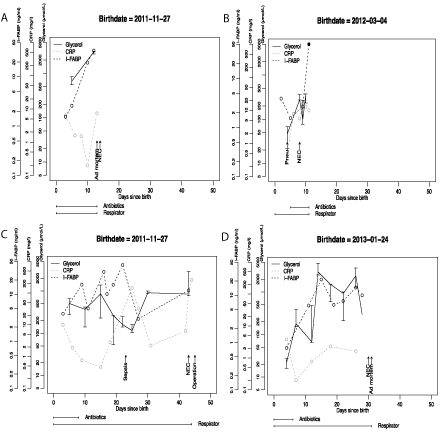
Figure 1
A-D. Glycerol, I-FABP and CRP levels in four infants developing NEC. Glycerol values are medians with bars for maximum and minimum values.
Figure 2
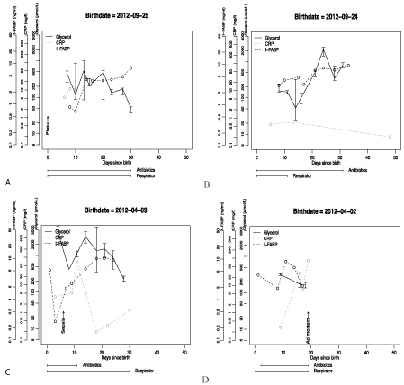
Figure 2
A-J. Glycerol, I-FABP and CRP levels in ten infants without clinical or radiological signs of NEC. Values for glycerol are medians with bars for maximum
and minimum values. Glycerol and I-FABP levels varied considerably during the observation periods, with both rising and falling concentrations at different time
points. Two infants (E, F) were operated for ligation of patent ductus arteriosus. K. One infant suffered from expansive intestinal necrosis due to mid-gut volvulus.
All infants were intubated and ventilation was maintained on respirator during the observation periods as indicated (Figure 1A-D, 2A-K). Three of them suffered from
pneumothorax due to ventilator trauma (Figure 1B, 2A, G). Antibiotic treatment was also initiated in all children, and maintained for different periods as indicated
(Figure 1A-D, 2A-K). Two of the infants displayed a significant persistent ductus arteriosus, and were operated on thereafter (Figure 2E, F). Severe infection and
sepsis was present in one patient (Figure 2C).
Materials and Methods
Study population and setting
The study was approved by the regional committee on medical
research ethics, and informed consent was obtained from the parents.
Preterm infants with a gestational age less than 28 weeks and
weighing <1500 g were included. No abdominal symptoms or other
clinical signs of illness were present in the infants, and there was
no evidence of any otherwise complicating disease on inclusion.
The infants were admitted to a level III neonatal intensive care
unit, and followed during a 4-week period. Any routine blood
testing and radiology scans were performed on a clinical basis. A
total of 15 infants were included during this period. Four of these
developed NEC stage 2 or 3, or NEC confirmed by histopathology.
The remaining 11 infants did not develop NEC during this period,
and therefore served as controls. The diagnosis of NEC was staged
according to a simplification of the Bell classification (Walsh and
Kliegman) [21], using 3 categories. Stage 1 was defined as suspected
NEC, with lethargy, abdominal distension, bloody stools or apnea.
Stage 2 was considered present when either X-rays or ultrasound
revealed pneumatosis intestinalis, portal gas, intestinal perforation
or ileus with dilated bowel loops. Stage 3 was defined when the
occurrence of organ failure was present in addition to the stage 2
criteria.
Microdialysis
The infants were monitored during a 4-week period, with
microdialysis measurements twice a week. All measurements
were performed using a clinically approved CMA 70 microdialysis
catheter (cut-off 20 kDa, 10 mm membrane length, Mdialysis
AB, Solna, Sweden). The microdialysis catheters were connected
to microinjection pumps (CMA 107, CMA Microdialysis AB,
Stockholm, Sweden) and perfused with an isotonic Ringer´s solution
with a flow rate of 1.0 µL/minute.
The microdialysis catheter was rectally inserted 10 mm, and
secured in position with tape. Initially, in situ stabilization was
allowed for 5 minutes, due to the non-traumatic placement of
the catheter. Microdialysate samples were then collected every 30
minutes for a total of 90 minutes. Samples were immediately put
into freezer at -20°C. Analyses of glucose, L-lactate, pyruvate and
glycerol were performed using an enzymatic colorimetric technique
on a CMA 600 Microdialysis Analyzer (CMA Microdialysis AB). The
CMA 600 Analyzer was automatically calibrated at start-up and recalibrated
every sixth hour using standard calibration solutions from
the manufacturer (CMA Microdialysis AB). Quality controls at two
different concentrations for each analysate were performed every
weekday. Total imprecision coefficient of variation was <10% for all
analysates.
Blood samples
Routine testing of blood samples included C-Rreactive Protein
(CRP) using the standard clinical laboratory method. Blood was
drawn for analysis of I-FABP. A volume of 100 µL in EDTA was
centrifuged, and plasma was stored at -70°C until analysis. I-FABP
was analyzed by a commercial sandwich-ELISA (DY3078, R&D
Systems, Minneapolis, MN, USA), in which a monoclonal antibody
specific for I-FABP was coated onto microtitre plates. Standards
and samples were pipetted into the wells and the peptide was bound
to the immobilized antibodies. After washing, a biotinylated antiI-FABP
antibody was added. Following incubation and washing, a
streptavidine-HRP conjugate was added to the wells. After incubation
and washing, a substrate solution was added. The development was
stopped and the absorbance was measured in a SpectraMax 250
(Molecular Devices, Sunnyvale, CA, USA). The concentrations in
the samples were determined by comparing the optical density of the
sample with the standard curve. The assays were calibrated against
highly purified recombinant human I-FABP. Measurements of
I-FABP were performed without knowledge of the clinical diagnoses.
Results
Microdialysis
Intraluminal microdialysate levels of glycerol were detectable.
However, the concentrations of lactate, glucose and pyruvate were
too low for analysis in all infants. During the observation periods,
the concentrations of glycerol, I-FABP and CRP varied considerably
in infants with NEC (Figure 1A-D). The mean levels of glycerol or
I-FABP at NEC diagnosis were not higher than before diagnosis, or
compared to controls.
The infants who did not develop NEC served as controls. In
these infants, both glycerol and I-FABP levels also revealed a high
degree of variation, with rising and falling concentrations during the
observation periods (Figure 2A-K). One infant developed symptoms
of intestinal ischemia but no signs of NEC on x-ray (Figure 2K).
Laparotomy revealed expansive intestinal necrosis, as a result of
mid-gut volvulus. In this patient, I-FABP clearly displayed an early
elevation, accompanied by a later rise of glycerol and CRP.
Discussion
Today, NEC is diagnosed by a combination of clinical, laboratory,
and radiological findings. These diagnostic methods lack high
specificity and sensitivity for NEC, especially in the early phase.
Certain indications of NEC are evident at a late stage during the
course of the disease, when intestinal necrosis is manifest. Early
diagnosis and treatment is important to reduce the morbidity and
mortality associated with NEC. Thus, there is a strong need for
improved diagnostic methods.
Microdialysis has previously been used to study intestinal
ischemia, both on humans and on rats. The advantage of the
microdialysis approach is that it measures metabolites of ischemia,
locally, in the organ of interest. In early stage experimental NEC, the
mucosa is primarily affected, as it is the most vulnerable part of the
gut wall [13]. This raises the opportunity to use intestinal intraluminal
microdialysis to identify anaerobic stress at an early stage, before
systemic levels of the metabolites are reached, and before the organ of
interest is severely damaged.
In our previous study on experimental intestinal ischemia
[12], signs of intestinal damage in the lumen were measured by
microdialysis, before systemic levels of the anaerobic metabolites
were reached. The intraluminal levels of glycerol also had a positive
correlation with aggravated histological mucosal damage. We
have previously studied intestinal intraluminal microdialysis in
experimentally induced early NEC in rat pups [13]. In the study,
elevated levels of glycerol and lactate were measured by placement of
a rectally inserted microdialysis catheter.
In humans, patients with recent abdominal surgery have been
monitored with microdialysis [22,23], and these studies have
demonstrated that microdialysis is a valuable tool for detecting
visceral ischemia using the intraperitoneal approach. However,
no clinical studies have previously been performed using the
intraluminal approach, partly because of the difficulty of placing the
microdialysis catheters in the gut lumen. In one study, subcutaneous
microdialysis was used in neonates that had recently undergone
surgery to monitor metabolic changes [24]. The rationale for using
the intraluminal approach instead of the intraperitoneal, is that
any early mucosal damage would be first detected in the gut lumen,
whereas any intraperitoneal detection of metabolites would reflect a
later stage in the disease.
To our knowledge this study is the first to apply intestinal
intraluminal microdialysis to detect hypoxic stress in extremely
preterm infants. Despite the low number of patients in this study,
we have found that intraluminal microdialysis of the bowel is easily
accessible by rectal placement of the microdialysis catheter, which
has the advantage of being a minimally invasive method. Although
it was noted that an elevation of glycerol was seen in infants with
NEC, it was not possible to detect any significant increase in glycerol
concentrations prior to clinical diagnosis. In the infants with other
complications than NEC, intraluminal glycerol values also varied
considerably, both rising and falling during the observation period.
Therefore it was not possible to differentiate between NEC and the
controls, by observing any increase in glycerol concentrations.
We have also been able to analyze plasma levels of I-FABP in
extremely preterm infants born before 28 weeks of gestation. Plasma
levels of I-FABP displayed a similar pattern as glycerol, with high
concentrations before the development of NEC, as well as in the
control infants. In a previous study of I-FABP as a diagnostic marker
of intestinal ischemia, suggested cut-off point for non-reversible
intestinal ischemia was 1.3 ng/ml [25]. A study on healthy preterm
infants with gestational age between 28 and 33 weeks, plasma
concentrations ranged between 0.46-4.5ng/ml [17]. An interesting
finding in our present study is that the concentrations of I-FABP, in
controls as well as infants with NEC, exceeded these levels even at an
early stage. These findings may suggest that the previous suggested
cut-off point or normality range of I-FABP is not relevant in this
patient category of extremely preterm infants. Infants with high
enterocyte turnover should theoretically display higher levels of
I-FABP. Another explanation could be that the high concentrations of
I-FABP in the present study reflect intestinal enterocyte damage. This
fact is supported by the high intraluminal concentrations of glycerol
at an early stage in controls as well as in infants later developing NEC.
It is highly valuable to establish knowledge regarding the normality
range of I-FABP levels in this patient category.
In the present pilot study, elevated intraluminal glycerol levels, as
well as plasma I-FABP, were detected in infants developing NEC as
well as in those who had no abdominal symptoms. Therefore, we were
not able to differentiate patients with NEC from control patients with
other diseases. Many of these controls, however, were severely ill, with
complications following extreme prematurity. Primarily, respiratoryand
ventilation-associated problems dominated, resulting in long
periods with low blood oxygen saturation levels. This relative hypoxic
state may result in a compromised oxygenation of the intestines. In
particular, the sensitive mucosal cell-layer might be affected, which
could result in a hypoxia-induced mucosal cell membrane decay
and release of glycerol and I-FABP into the intestinal lumen. Other
complications and diseases as sepsis, infections, persistent ductus
arteriosus, and anemia were also present in the control group, which
aggravates the intestinal distress.
A potential methodological problem of microdialysis is that
it only measures a relative concentration of the metabolites in the
compartment of interest. This fact makes it difficult to compare the
absolute values of two different measurements performed at different
time intervals. To overcome the problem of relative concentrations,
ratios such as the lactate/pyruvate ratio are often used. The lactate/
pyruvate ratio is considered to be independent of changes in relative
recovery, making it a useful quantitative measure [18]. In this study
however, intraluminal levels of lactate and pyruvate were too low to
be measured, regardless of NEC or not. The lactate/pyruvate ratio,
therefore, could not serve as an indicator of hypoxic damage in the
intestines in this setup. The concentrations of glycerol, on the other
hand, were much higher but varied considerably during the course
of the 4-week observation period. A higher relative recovery could
be achieved by using microdialysis catheters with longer membranes
or by using a lower perfusion flow rate. This theoretically results in
higher concentrations of lactate and pyruvate, enabling calculation
of the lactate/pyruvate ratio. Initially, we tried to use a 30 mm
membrane, but it was not possible due to the anatomical limitations
of the extremely preterm infants. A lower perfusate flow rate would
also increase the recovery, but this was not practically possible in the
set up.
In conclusion, this preliminary study has shown that rectal
intraluminal microdialysis is safe and could provide a valuable noninvasive
aid to detect hypoxia-induced intestinal damage or ischemic
stress in extremely preterm infants. However, it was not possible
to predict or differentiate NEC from other diagnoses, by detecting
elevated levels of glycerol or I-FABP.
Figure 2
Figure 2
A-J. Glycerol, I-FABP and CRP levels in ten infants without clinical or radiological signs of NEC. Values for glycerol are medians with bars for maximum
and minimum values. Glycerol and I-FABP levels varied considerably during the observation periods, with both rising and falling concentrations at different time
points. Two infants (E, F) were operated for ligation of patent ductus arteriosus. K. One infant suffered from expansive intestinal necrosis due to mid-gut volvulus.
All infants were intubated and ventilation was maintained on respirator during the observation periods as indicated (Figure 1A-D, 2A-K). Three of them suffered from
pneumothorax due to ventilator trauma (Figure 1B, 2A, G). Antibiotic treatment was also initiated in all children, and maintained for different periods as indicated
(Figure 1A-D, 2A-K). Two of the infants displayed a significant persistent ductus arteriosus, and were operated on thereafter (Figure 2E, F). Severe infection and
sepsis was present in one patient (Figure 2C).
Figure 2
Figure 2
A-J. Glycerol, I-FABP and CRP levels in ten infants without clinical or radiological signs of NEC. Values for glycerol are medians with bars for maximum
and minimum values. Glycerol and I-FABP levels varied considerably during the observation periods, with both rising and falling concentrations at different time
points. Two infants (E, F) were operated for ligation of patent ductus arteriosus. K. One infant suffered from expansive intestinal necrosis due to mid-gut volvulus.
All infants were intubated and ventilation was maintained on respirator during the observation periods as indicated (Figure 1A-D, 2A-K). Three of them suffered from
pneumothorax due to ventilator trauma (Figure 1B, 2A, G). Antibiotic treatment was also initiated in all children, and maintained for different periods as indicated
(Figure 1A-D, 2A-K). Two of the infants displayed a significant persistent ductus arteriosus, and were operated on thereafter (Figure 2E, F). Severe infection and
sepsis was present in one patient (Figure 2C).
References
- Patel BK, Shah JS. Necrotizing enterocolitis in very low birth weight infants: a systemic review. ISRN gastroenterol. 2012; 2012: 562594.
- Henry MC, Moss RL. Necrotizing enterocolitis. Annu Rev Med. 2009; 60: 111-124.
- Lin PW, Stoll BJ. Necrotising enterocolitis. Lancet. 2006; 368: 1271-1283.
- Neu J, Walker WA. Necrotizing enterocolitis. N Engl J Med. 2011; 364: 255-264.
- Petrosyan M, Guner YS, Williams M, Grishin A, Ford HR. Current concepts regarding the pathogenesis of necrotizing enterocolitis. Pediatr Surg Int. 2009; 25: 309-318.
- Fox TP, Godavitarne C. What really causes necrotising enterocolitis? ISRN gastroenterology. 2012; 2012: 628317.
- Emmertsen KJ, Wara P, Soerensen FB, Stolle LB. Intestinal microdialysis--applicability, reproducibility and local tissue response in a pig model. Scand J Surg. 2005; 94: 246-251.
- Solligard E, Juel IS, Bakkelund K, Johnsen H, Saether OD, Gronbech JE, et al. Gut barrier dysfunction as detected by intestinal luminal microdialysis. Intensive Care Med. 2004; 30: 1188-1194.
- Solligard E, Juel IS, Bakkelund K, Jynge P, Tvedt KE, Johnsen H, et al. Gut luminal microdialysis of glycerol as a marker of intestinal ischemic injury and recovery. Crit Care Med. 2005; 33: 2278-2285.
- Sommer T. Microdialysis of the bowel: the possibility of monitoring intestinal ischemia. Expert Rev Med Devices. 2005; 2: 277-286.
- Sommer T, Larsen JF. Detection of intestinal ischemia using a microdialysis technique in an animal model. World J Surg. 2003; 27: 416-420.
- Hogberg N, Carlsson PO, Hillered L, Meurling S, Stenback A. Intestinal ischemia measured by intraluminal microdialysis. Scand J Clin Lab Invest. 2012; 72: 59-66.
- Hogberg N, Carlsson PO, Hillered L, Stenback A, Lilja HE. Intraluminal intestinal microdialysis detects markers of hypoxia and cell damage in experimental necrotizing enterocolitis. J Pediatr Surg. 2012; 47: 1646-1651.
- Kanda T, Fujii H, Tani T, Murakami H, Suda T, Sakai Y, et al. Intestinal fatty acid-binding protein is a useful diagnostic marker for mesenteric infarction in humans. Gastroenterology. 1996; 110: 339-343.
- Lieberman JM, Sacchettini J, Marks C, Marks WH. Human intestinal fatty acid binding protein: report of an assay with studies in normal volunteers and intestinal ischemia. Surgery. 1997; 121: 335-342.
- Edelson MB, Sonnino RE, Bagwell CE, Lieberman JM, Marks WH, Rozycki HJ. Plasma intestinal fatty acid binding protein in neonates with necrotizing enterocolitis: a pilot study. J Pediatr Surg. 1999; 34: 1453-1457.
- Guthmann F, Borchers T, Wolfrum C, Wustrack T, Bartholomaus S, Spener F. Plasma concentration of intestinal- and liver-FABP in neonates suffering from necrotizing enterocolitis and in healthy preterm neonates. Mol Cell Biochem. 2002; 239: 227-234.
- Derikx JP, Evennett NJ, Degraeuwe PL, Mulder TL, van Bijnen AA, van Heurn LW, et al. Urine based detection of intestinal mucosal cell damage in neonates with suspected necrotising enterocolitis. Gut 2007; 56: 1473-1475.
- Evennett NJ, Hall NJ, Pierro A, Eaton S. Urinary intestinal fatty acidbinding protein concentration predicts extent of disease in necrotizing enterocolitis. J Pediatr Surg. 2010; 45: 735-740.
- Thuijls G, Wijck K, Grootjans J, Derikx JP, van Bijnen AA, Heineman E, et al. Early diagnosis of intestinal ischemia using urinary and plasma Fatty Acid binding proteins. Ann Surg 2011; 253: 303-308.
- Walsh MC, Kliegman RM. Necrotizing enterocolitis: treatment based on staging criteria. Pediatr Clin North Am. 1986; 33: 179-201.
- Jansson K, Redler B, Truedsson L, Magnuson A, Ungerstedt U, Norgren L. Postoperative on-line monitoring with intraperitoneal microdialysis is a sensitive clinical method for measuring increased anaerobic metabolism that correlates to the cytokine response. Scand J Gastroenterol. 2004; 39: 434-439.
- Jansson K, Ungerstedt J, Jonsson T, Redler B, Andersson M, Ungerstedt U, et al. Human intraperitoneal microdialysis: increased lactate/pyruvate ratio suggests early visceral ischaemia. A pilot study. Scand J Gastroenterol. 2003; 38: 1007-1011.
- Hildingsson U, Sellden H, Ungerstedt U, Marcus C. Microdialysis for metabolic monitoring in neonates after surgery. Acta Paediatr. 1996; 85: 589-594.
- Vermeulen Windsant IC, Hellenthal FA, Derikx JP, Prins MH, Buurman WA, Jacobs MJ, et al. Circulating intestinal fatty acid-binding protein as an early marker of intestinal necrosis after aortic surgery: a prospective observational cohort study. Ann Surg. 2012; 255: 796-803.