Case Report
Severe Bilateral Degenerative Temporomandibular Joint Disease with Erosion into the Middle Cranial Fossa: An Uncommon Etiology
Dolwick MF*, Schlott BJ, McPherson TR and Holman MR
Department of Oral and Maxillofacial Surgery, University of Florida College of Dentistry, USA
*Corresponding author: M. Franklin Dolwick, Department of Oral and Maxillofacial Surgery, University of Florida College of Dentistry, USA
Published: 16 Aug, 2016
Cite this article as: Dolwick MF, Schlott BJ, McPherson TR, Holman MR. Severe Bilateral Degenerative Temporomandibular Joint Disease with Erosion into the Middle Cranial Fossa: An Uncommon Etiology. Clin Surg. 2016; 1: 1083.
Abstract
Erosion of the temporomandibular joint into the middle cranial fossa due to severe degenerative
joint disease is an extremely rare occurrence. There have been multiple cases of the condyle being
displaced into the middle cranial fossa as a result of trauma and pathology. The patient in this
case report presented with erosive arthropathy of the bilateral temporomandibular joints with
nearly complete loss of the condylar heads which were bilaterally displaced into the middle cranial
fossa with dural thickening. Treatment options were complicated by partial edentulism and the
potential for intracranial complications. The patient was reconstructed with bilateral TMJ Concepts
prostheses using her dentures as a guide for establishing the appropriate vertical dimension.
Keywords: Middle cranial fossa; Joint disease
Introduction
Displacement of a condyle into the middle cranial fossa is a relatively rare occurrence generally
having a traumatic or pathologic etiology [1,2]. There have also been cases reported as post-operative
complications resulting in an alloplastic prosthesis eroding into the middle cranial fossa with a
resulting cerebrospinal fluid leak [3].
Severe degenerative joint disease is characterized by pain, limited opening, deformity of the
articular disc, closed lock, crepitus, and degenerative osseous changes in the condyle or fossa.
Approximately five percent of patients who suffer from temporomandibular disorders require
surgical management [4]. Patients requiring surgical management generally have undergone
multiple procedures and some ultimately require reconstruction with a prosthesis to address
functional deficits. The patient we are presenting had an unusual presentation, chief complaint, and
etiology. She had no prior surgical or non-surgical intervention for her severe bilateral degenerative
joint disease and erosion into the middle cranial fossa bilaterally.
Case Report
A 69 year old female with a past medical history significant for renal artery stenosis, COPD,
polyarticular osteoarthritis, and acute pancreatitis of which the underlying etiology was unclear
presented with the complaint of progressively worsening retrognathia and malocclusion. Her chief
complaint was primarily centered around functional status, not pain. She began to notice changes in
her occlusion initially and reported difficulty incising and chewing food. The patient had a skeletal
class II appearance with a progressively worsening anterior open bite. She was edentulous in the
maxilla, partially edentulous in the mandible with retention of the mandibular anterior teeth from
22-28, and had apertognathia. The distance from the maxillary alveolus to mandibular incisors
on maximal opening was 30mm. She had mild reproducible pain on palpation of the bilateral
temporomandibular joint capsule. Neurologically she was intact.
Diagnostic imaging
The patient underwent computed tomography of the head which demonstrated an erosive
arthropathy of bilateral temporomandibular joints with nearly complete loss of the condylar heads,
expansion and thickening of the joint capsule, and marked thinning and remodeling of the condylar
fossa (Figure 1). There was bone dehiscence superiorly bilaterally in the condylar fossa with dural
thickening which was greater on the right. Given these findings we obtained a contrasted stealth MRI
for further evaluation of the middle cranial fossa and surgical planning (Figure 2). The MRI findings
were consistent with the prior CT head. The MRI demonstrated
bulging of the expanded temporomandibular joint capsule into the
middle cranial fossa with dural thickening bilaterally (Figure 1 and 2).
Treatment
There has been multiple treatment algorithms described for
displacement of a condyle into the middle cranial fossa with a
traumatic etiology which involve closed reduction, open reduction
with craniotomy, and condylotomy [5]. Due to the etiology of
erosion in our patient there was nearly complete loss of the condylar
head. Treatment options were discussed with the patient and she
elected to undergo bilateral total joint replacement to restore her
acquired skeletal malocclusion and vertical dimension in an effort
to improve her function. Treatment planning was complicated
by the potential for an intra-operative cerebrospinal fluid leak and
intracranial hemorrhage [6]. There have been multiple cases of a
condyle displaced into the middle cranial fossa due to other etiologies
one of which an alternative surgical approach was used which
involved leaving the condyle in the middle cranial fossa predisposing
the patient to less risk of intracranial hemorrhage [7]. Given the
displacement of the remaining condyles into the middle cranial fossa
the patient was referred to the neurosurgery department for a preoperative
evaluation. Neurosurgery agreed to be available for intraoperative
assistance and consented the patient for a sub temporal
craniotomy, fascia latta, fat graft, and lumbar drain placement if
indicated. Preoperatively alginate impressions were taken and her
existing dentures were duplicated. The casts and duplicated dentures
were used to establish the occlusion and vertical dimension. This
along with imaging was sent to TMJ Concepts for construction of the
bilateral total joint prostheses (Figure 3).
The patient was taken to the operating room and underwent
nasotracheal intubation. After positioning and prepping the patient
her left joint space was approached from a preauricular incision and
the left mandible from a submandibular approach. A 701 fissure bur
was used to make an osteotomy and remove the remaining portion of
the condylar head. On exploration of the glenoid fossa an extensive
amount of granulation tissue was found and removed. There was
no cerebrospinal fluid leak observed and the dura remained intact.
The mandible was pushed superiorly from the submandibular
approach and an osteotomy was made from the sigmoid notch to
the posterior aspect of the mandible to accommodate the alloplastic
joint. The temporal bone was contoured with a bur to accommodate
the fossa component then the fossa component was secured. An
identical approach was made to the right joint space and mandible
with similar osteotomies and removal of granulation tissue. The
fossa component was fitted in a similar fashion as the contralateral
side. The duplicated dentures were placed in the mouth along with
Karlis screws. The patient was placed into maxillomandibular fixation
to establish the proper vertical dimension. Once the occlusion
was established the condylar portion bilaterally was temporarily
secured with two bicortical screws and the patient was released from
maxillomandibular fixation to verify that the occlusion was correct,
stable, and repeatable. The condylar portions were then permanently
fixated with bicortical screws. An abdominal fat graft was harvested
from the umbilicus region and grafted bilaterally to the condylar
region. All surgical sites were then irrigated and closed. The bilateral
condylar heads and associated soft tissue were sent as specimens. The
diagnosis on the final pathology report was fibro cartilaginous tissue,
granulation tissue, and associated cortical bone.
Postoperatively the patient did well and was sent home on the
diet of her choice on postoperative day two. She was seen for follow
up at two and four week intervals and was recovering appropriately
without complication. At four weeks postoperatively she opened to a
maximum incisal opening with her previous dentures to a distance of
31mm and was consuming a regular diet. (See figure 4) She was lost
to follow up after four weeks.
Figure 1
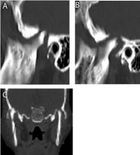
Figure 1
Computed axial tomography of the head. A) Oblique sagittal view
of the right condylar head. B) Oblique sagittal view of the left condylar head.
C: Coronal view of the bilateral condylar heads.
Figure 2
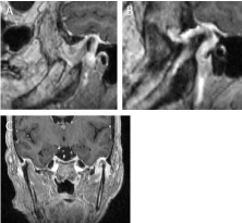
Figure 2
MRI of the brain with intravenous contrast. A) Oblique sagittal view of the right condylar head. B) Oblique sagittal view of the left condylar head.
C: Coronal view of the bilateral condylar heads.
Figure 3
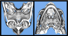
Figure 3
Three dimensional reconstruction of the head CT. A) Superior view
demonstrating erosion of the condylar head into the middle cranial fossa. B)
Inferior view of the mandible.
Figure 4
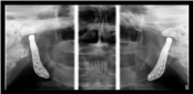
Figure 4
Panoramic radiograph, Postoperative panoramic radiograph.
Conclusion
While temporomandibular disorders are common among the population most are managed conservatively with only five percent requiring surgical intervention [4]. Most patients who undergo surgical management with a bilateral temporomandibular joint replacement with an alloplastic prosthesis have exhausted all other surgical options for severe degenerative joint disease. Bilateral native temporomandibular joint erosion into the middle cranial fossa is an extremely rare occurrence. It is also extremely rare for a patient to present at such an advanced stage in the disease process with minimal symptoms and requiring bilateral total joint replacement as the primary intervention. When the condyle is displaced into the middle cranial fossa the etiology is generally a result of trauma or pathology, other etiologies include advanced aging, malignancy, and condromatosis of the temporomandibular joint.
References
- Lee T, Green R, Hsu J. Central condylar displacement with brain abscess from chronic mandibular osteomyelitis. The Laryngoscope. 2013; 123: 1369–1373.
- Vaezi T, Rajaei SA, Abrishami MH, Taghvaei ME. Dislocation of the mandibular condyle into the middle cranial fossa: a case report. Oral and maxillofacial surgery. 2014; 18: 69-73.
- Chuong R, Piper MA. Cerebrospinal fluid leak associated with proplast implant removal from the temporomandibular joint. Oral surgery, oral medicine. Oral pathology. 1994; 74: 422-425.
- Dolwick MF. Temporomandibular joint surgery for internal derangement. Dental clinics of North America. 2007; 51: 195-208.
- Y. He, Zhang Y, Li ZL, An JG, Yi ZQ, Bao SD. Treatment of traumatic dislocation of the mandibular condyle into the cranial fossa: velopment of a probable treatment algorithm. International journal of oral and maxillofacial surgery. 2015; 44: 864-870.
- Struewer J, Kiriazidis I, Figiel J, Dukatz T, Frangen T, Ziring E. Dislocation of the mandibular condyle into the middle cranial fossa causing an epidural haematoma. J Craniomaxillofac Surg. 2012; 40: 396-399.
- Guevara HG, Gavranich J, Moreira TA, Vasconcellos V, Leandro LL. Temporomandibular joint prostheses: An alternative for impacted mandibular condyle in middle cranial fossa. Revista Española de Cirugía Oral y Maxilofacial. 2013; 35: 181-185.