Case Report
Use of Panniculus Flap with Delayed Penile Reconstruction for Management of Buried Penis in a Patient with Massive Localized Lymphedema of the Scrotum: A Case Report
Azouz V, Kareh A and Bialowas C*
Department of Surgery, University of Texas Health Science Center, USA
*Corresponding author: Christie Bialowas, Department of Surgery, Division of Plastic Surgery, The University of Texas Health Science Center at San Antonio, 7703 Floyd Curl Drive, San Antonio, TX 78229-3900, USA
Published: 13 Aug, 2016
Cite this article as: Azouz V, Kareh A, Bialowas C. Use of Panniculus Flap with Delayed Penile Reconstruction for Management of Buried Penis in a Patient with Massive Localized Lymphedema of the Scrotum: A Case Report. Clin Surg. 2016; 1: 1081.
Abstract
Buried penis is a rare debilitating phenomenon that has been associated with obesity, lymphedema,
radical circumcision, and penoscrotal elephantiasis. Traditional management of this condition
involves limited panniculectomy, excision of penile shaft skin, and skin grafting. Buried penis
secondary to massive localized lymphedema of the scrotum is seldom described in the literature and
further complicates the use of local reconstruction as local tissue is damaged and may be unfit for
reconstructive purposes. The authors report the case of a morbidly obese patient with buried penis
secondary to massive localized lymphedema of the scrotum reconstructed using a panniculus flap
and delayed penile skin grafting. After undergoing scrotal reconstruction using a panniculus flap,
the patient returned to the operating room five days later to undergo penile reconstruction with
meshed split thickness skin grafting. Sequential compression wrapping between the first and second
procedures allowed the swelling and edema to decrease significantly so that the penile bed would
be more amenable to split thickness skin graft. In treating buried penis in the setting of massive
scrotal lymphedema, the authors propose reconstructive staging and sequential compression
dressings between procedures to maximize patient safety by limiting postoperative complications
and minimize edema.
Keywords: Buried penis; Massive localized lymphedema; Scrotal reconstruction
Introduction
Acquired buried penis is a rare phenomenon that has been associated with obesity, lymphedema,
radical circumcision, and penoscrotal elephantiasis. Those afflicted by the rare condition suffer
from sexual dysfunction, psychological stress, difficulty ambulating, surgical infections, and chronic
wound ulcerations. Surgically correcting a buried penis is a unique and difficult challenge that
requires a multidisciplinary approach including plastic and urologic surgeons.
Buried penis secondary to massive localized lymphedema of the scrotum is seldom described
in the literature and further complicates the use of local reconstruction as local tissue is damaged
and may be unfit for reconstructive purposes [1]. Careful preoperative planning that addresses this
multifactorial pathology is imperative to ensure appropriate treatment and recovery. We report the
case of a morbidly obese patient with buried penis secondary to massive localized lymphedema of
the scrotum reconstructed using a panniculus flap and delayed penile skin grafting. We emphasize
the importance of staging operative procedures in morbidly obese patients to optimize patient safety
by reducing intraoperative and postoperative complications.
Materials and Methods
A 44 year-old morbidly obese male (BMI 71) after 100lbs weight loss presented with massive
lymphedema and ulcerations of his mons and scrotum that had significantly enlarged over the past
three years. On exam his scrotum was significantly inflamed, making it difficult for him to ambulate
(Figure 1 and 2). Due to the extent of his buried penis, the patient voided by allowing urine to
dribble from the area between his pannus and scrotum for over 14 years. This made it difficult
for him to have proper hygiene and impaired him from engaging socially. He agreed to undergo
reconstructive surgery with the plastic surgery and urology team to treat this debilitating condition.
In the operative theater the patient underwent mons lipectomy and scrotal resection with local
tissue rearrangement in collaboration with urology who performed a circumcision and cystoscopy.
Prior to scrotal resection, urology identified the penis and performed
a dorsal slit procedure that extended 20cm onto and through the
scrotal skin until the glans was identified. The penile shaft was noted
to be denuded and the scrotal skin had invaginated. The surrounding
scrotal tissue was then dissected using sharp and blunt dissection,
liberally using cautery, clips, suture ligasures, and the Ligasure device.
The total excised scrotal tissue was measured and weighed 35 pounds.
The phallus was then dissected free to mobilize the penile shaft and
the suspensory ligament.
To reconstruct the scrotum the superior portion of the mons
was taken down and tacked to the pubis and the base of the penis.
Next two 10 x 20 cm flaps from the residual pannus were cleared of
lymphedematous fluid, folded over and reapproximated to create a
neoscrotum. This was then tacked up to the base of the penis laterally
on each side to allow the penis to be up against the pubis. At this time
we were approaching our five hour safety threshold for the procedure
and deferred skin grafting. The patient’s prolonged surgical course
in the lithotomy increased his risk for rhabdomyolysis, neuropraxia
and compartment syndrome [2-4]. By staging the reconstruction
we were also able to avoid possible complications of seroma and
infection secondary to swelling [5]. The patient was then transferred
to the PACU in stable condition. The length of the procedure was just
under five hours.
Figure 1
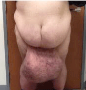
Figure 1
Text Here.
Figure 2
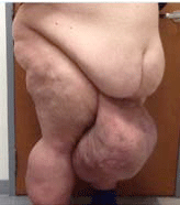
Figure 2
Text Here.
Results
On postoperative day 2 the patient endorsed ambulating and adequate pain control. After two additional days of observation, the patient was discharged home with the foley in place. Five days later, the patient returned to the operative room for delayed penile reconstruction with split thickness skin graft. Intraoperatively, the patient’s scrotal edema was noted to be markedly decreased, the penile shaft was then skin grafted and subsequently dressed with bacitracin and Xeroform. After a bolster was placed and stapled in place to the body and glans of the penis, the donor site was dressed in the usual fashion and the patient was transferred to PACU in stable condition. The estimated blood loss was 10ml and the procedure length was 1.5 hours. The patient’s postoperative course was uncomplicated and he was discharged the next day. The sequential scrotal wrapping controlled the edema and produced an appropriately sized scrotum (Figure 3 and 4). The patient can now wear pants and can walk several blocks which were impossible before the procedure.
Figure 3
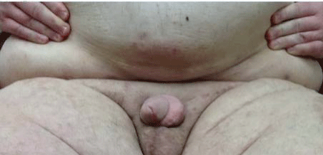
Figure 3
Text Here.
Figure 4
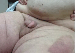
Figure 4
Text Here.
Discussion
Though the management of buried penis has been extensively
described in the literature Pestana et al. [5] are first to present a
modified algorithm to the management of buried penis [6-10]. Their
approach initially involves releasing the scar contracture of the shaft
skin, resecting skin between the panniculus fold and pubic hair if
there is abdominal fat pad impingement, and then releasing the
suspensory ligament if more phallus exposure is required. Primary
closure is then pursued if there is adequate shaft skin and tissue for
coverage of the penis, but in most cases this is not possible due to the
high likelihood that shaft skin removal is required as part of the initial
release. In these cases, a split thickness skin graft is recommended
for coverage with tissue flaps reserved as a secondary option [6,7,9].
While the algorithm proposed by Pestana et al. addresses buried penis
management, it does not address operative management required
in patients with buried penis in the setting of massive localized
lymphedema of the scrotum.
Massive Localized Lymphedema (MLL), the condition of
chronically impaired lymphatic drainage originally coined by Farshid
and Weiss, appears to be increasing secondary to the rise in the
incidence of morbid obesity in today’s society [11-16]. The chronic
retention of fluid in the extravascular space expands connective
tissue causing loss of elasticity and inflammation. This destructive
process is exacerbated by the decreased inflammatory cell flow which
predisposes the tissue to infection. For these reasons, afflicted tissue
may be unfit for local reconstruction.
Conventional non-operative management of MLL consists of
compression stockings, elevation, and bed rest has been deemed
ineffective in treating scrotal MLL, especially when activities of
daily living are impaired [13,17]. Surgical treatment options for
MLL include physiologic and ablative reconstruction operations.
Physiologic reconstruction involves building channels that expand
the transport capacity of lymphatic fluid. This technically challenging
option has yet to be established in the treatment of scrotal MLL.
Ablative reconstruction involves removing effected excess scrotal skin
and soft tissue either minimally or entirely. Minimal skin resection
and soft tissue resection may allow for primary closure while more
aggressive resection may necessitate resurfacing with skin grafting
[9,18]. Although Full Thickness Skin Grafts (FTSG) have traditionally
been used for penile reconstruction, successful use of Split Thickness
Skin Graft (STSG) has been extensively described in the literature
[7,8,19-21]. STSG is ideal due to high rates of success in contaminated
fields, ease of harvest, and excellent cosmetic results [19]. Due to the
high risk of contamination and difficult shape of the graft bed, we
elected to use meshed STSG in our case.
At the conclusion of the first case, the patient’s reconstructed
scrotum was wrapped with compression dressings in order to control
the amount of lymphedema, wound breakdown, and swelling to the
area. Daily sequential wrapping of the scrotum was performed until
the patient was taken back to the OR. This decision was based on the
standard conservative management of lymphedema to the scrotum,
which consists of a penile compression bandage and tight scrotal
support [22]. The extended period of time between reconstruction
and STSG in addition to compression with Ace bandages allowed
the penile tissue to be more favorable for STSG due to decreased
swelling and lymphedema to the graft bed. This interval time period
also allowed for early ambulation which helped minimize the
patient's risk for postoperative Deep Venous Thrombosis (DVT). Our
patient’s Caprini Score was calculated as 12 (Age >41, major surgery
>3 hours, BMI >50, hx of DVT), which is considered high risk for
the development of DVTs [23]. Due to his multiple risk factors, the
classically described period of approximately 4 days of immobility
and bed rest to ensure good penile graft take was not observed [19].
In our case, we did not encounter any problems with the graft despite
early ambulation, leading us to conclude that as long as the graft is
bolstered and dressed properly bed rest is not necessary for the graft
to take successfully.
Staging the reconstruction not only helped limit edema and
postoperative complications, it also maximized patient safety. Due to
the patient's distorted anatomy, scrotal dissection required meticulous
technique as multiple large vessels that resembled spermatic cord
were encountered. As reported in the urologic surgery literature,
obese patients undergoing surgery lasting five hours or longer in the
lithotomy and exaggerated lithotomy position are at increased risk
of risk of rhabdomyolysis, subsequent renal injury, compartment
syndrome and neuropraxia. [2-4,24] Our patient’s morbid obesity and
prolonged operative time put him at high risk for developing these
complications. By staging the operative procedures, we were able
to reduce his intraoperative and postoperative risks. Therefore, we
encourage staging of these types of prolonged procedures especially
in patients whose BMI is greater than 50.
Conclusion
Lymphedema of the penis can be debilitating. It causes lymph obstruction, uncomfortable swelling and inflammation, and impairment of daily activities. Buried penis secondary to massive scrotal lymphedema is a unique reconstructive challenge that should be addressed with staged procedures to limit edema, postoperative complications, and to maximize patient safety.
References
- Halperin TJ, Slavin SA, Olumi AF, Borud LJ. Surgical Management of Scrotal Lymphedema Using Local Flaps. Ann Plast Surg. 2007; 59: 67-72.
- Guella A, Al Oraifi I. Rhabdomyolysis and acute renal failure following prolonged surgery in the lithotomy position. Saudi J Kidney Dis Transpl. 2013; 24: 330-332.
- Vijay MK, Vijay P, Kundu AK. Rhabdomyolysis and myogloginuric acute renal failure in the lithotomy/exaggerated lithotomy position of urogenital surgeries. Urol Ann. 2011; 3: 147–150.
- Targa L, Droghetti L, Caggese G, Zatelli R, Roccella P. Rhabdomyolysis and operating position. Anaesthesia. 1991; 46: 141–143.
- Warren AG, Peled ZM, Borud LJ. Surgical correction of a buried penis focusing on the mons as an anatomic unit. J Plast Reconstr Aesthet Surg. 2009; 62: 388–392.
- Pestana IA, Greenfield JM, Walsh M, Donatucci CF, Erdmann D. Management of “Buried” Penis in Adulthood: An Overview. Plast Reconstr Surg. 2009; 124: 1186–1195.
- Tang S-H, Kamat D, Santucci RA. Modern Management of AdultAcquired Buried Penis. Urology. 2008; 72: 124–127.
- Figler BD, Chery L, Friedrich JB, Wessells H, Voelzke BB. Limited Panniculectomy for Adult Buried Penis Repair. Plast and Reconstr Surg. 2015; 136: 1090–1092.
- Westerman ME, Tausch TJ, Zhao LC, Siegel JA, Starke N, Klein AK, et al. Ventral Slit Scrotal Flap: A New Outpatient Surgical Option for Reconstruction of Adult Buried Penis Syndrome. Urology. 2015; 85: 1501–1504.
- Shapiro SR. Surgical treatment of the “buried” penis. Urology. 1987; 30: 554–559.
- Farshid G, Weiss SW. "Massive Localized Lymphedema in the Morbidly Obese". Am J Surg Pathol. 1998; 22: 1277-1283.
- Chopra K, Tadisina KK, Brewer M, Holton LH, Banda AK, Singh DP. Massive Localized Lymphedema Revisited: A Quickly Rising Complication of the Obesity Epidemic. Ann Plast Surg. 2015. 74: 126–132.
- Champaneria MC, Workman A, Kao H, Ray AO, Hill M. Reconstruction of massive localized lymphoedema of the scrotum with a novel fasciocutaneous flap: A rare case presentation and a review of the literature. J Plast Reconstr Aesthet Surg. 2013; 66: 281–286.
- Asch S, James WD, Castelo-Soccio, L. Massive localized lymphedema: An emerging dermatologic complication of obesity. J Am Acad of Dermatol. 2008; 59: S109–S110.
- Evans RJ, Scilley C. Massive localized lymphedema: A case series and literature review. Can J Plast Surg. 2011; 19: e30–e31.
- Lee S, Han JS, Ross HM, Epstein JI. Massive localized lymphedema of the male external genitalia: a clinicopathologic study of 6 cases. Hum Pathol. 2013; 44: 277–281.
- Machol IV JA, Langenstroer P, Sanger JR. Surgical reduction of scrotal massive localized lymphedema (MLL) in obesity. J Plast Reconstr Aesthet Surg. 2014; 67: 1719–1725.
- Otsuki Y, Yamada K, Hasegawa K, Kimata Y, Suami H. Overview of Treatments for Male Genital Lymphedema: Critical Literature Review and Anatomical Considerations. Plast Reconstr Surg. 2012; 129: 767e–769e.
- Alwaal A, McAninch JW, Harris CR, Breyer BN. Utilities of SplitThickness Skin Grafting for Male Genital Reconstruction. Urology. 2015; 86: 835–839.
- Singh V, Sinha RJ, Sankhwar SN, Kumar V. Reconstructive Surgery for Penoscrotal Filarial Lymphedema: A Decade of Experience and Follow-up. Urology. 2011; 77: 1228–1231.
- Black PC, Friedrich JB, Engrav LH, Wessells H. Meshed Unexpanded Split-Thickness Skin Grafting for Reconstruction of Penile Skin Loss. J Urol. 2004; 172: 976–979.
- Garaffa G, Christopher N, Ralph DJ. The management of genital lymphoedema. BJU Int. 2008; 102: 480–484.
- Pannucci CJ, Bailey SH, Dreszer G, Fisher C, Zumsteg JW, Jaber RM, et al. Validation of the Caprini Risk Assessment Model in Plastic and Reconstructive Surgery Patients. J Am Coll Surg. 2011; 212: 105–112.
- Kikuno N, Urakami S, Shigeno K, Kishi H, Shiina H, Igawa M. Traumatic rhabdomyolysis resulting from continuous compression in the exaggerated lithotomy position for radical perineal prostatectomy. Int J Urol. 2002; 9: 521–524.