Case Report
Spindle Cell Carcinoma of the Lower Limbs: A Case Report
Niu F1*, Lai W2, Fu Q1, Ma G2 and Gao Y1
1Department of Orthopedics, Yangzhou Hospital of Traditional Chinese Medicine, China
2Department of Orthopedics, Xinyuan Hospital of Traditional Chinese Medicine, China
*Corresponding author: Feng Niu, Department of Orthopedics, Yangzhou Hospital Of Traditional Chinese Medicine, 577 Wenchang West Street, Yangzhou, Jiangsu, 225002, P.R. China
Published: 23 Jul, 2016
Cite this article as: Niu F, Lai W, Fu Q, Ma G, Gao Y. Spindle Cell Carcinoma of the Lower Limbs: A Case Report. Clin Surg. 2016; 1: 1074.
Abstract
Spindle cell carcinoma of the leg is an unusual tumor. It is important to increase the recognition of the disease and improve the level of clinical diagnosis. The current study presents a case
of spindle cell carcinoma with clinical, imaging and pathological examination. A 65-year-old
female presented to the Xinyuan Hospital of Traditional Chinese Medicine suffering from a mass
about 3.6 × 1.6 × 15.0cm on the back skin of her right leg for approximately one month. Imaging
examination by leg computed tomography scan revealed a 9.2 × 4.0 cm solid mass infiltrated into
the subcutaneous fat in the back skin of the right tibia. The patient underwent resection of the mass
on March, 17, 2013. Patholocical examination showed the tumor was composed of small, elongated
cords in a tightly packed arrangement. Tumor cells were smaller and cube-shaped or oval and lowgrade
nuclei. Occasionally, necrosis and foam cell infiltration were observed. The tumor reappeared
after one year later, a 6.0 × 7.0 × 15.0 cm mass appeared on the incision with bleeding after slight
impact. The patient underwent resection of the recurrence tumor and VSD covered the defect of
the skin on July, 10, 2014. The tumor reappeared again after 10 days and bleeded combined necrosis
and foul smell. Continuing bleeding cause the hypoproteinemia and emaciation for 3 months. The
patient cannot endure the miserable experience and asked amputation of the knee on Oct, 16, 2014.
However, a new mass about 1.6 × 1.6 × 1.0 cm was found in the skin of the popliteal fossa. Patholocical
examination showed the new mass was spindle cell carcinoma. However, subsequent chemotherapy,
radiation and immunohistochemical markers weren’t underwent because of the patient’s personal
economic reason. The patient is still alive after 7 months without evidence of disease, we don’t
receive the recurrence report after the follow investigation until now.
Keywords: Spindle cell carcinoma; Lower limbs; Skin
Introduction
Spindle cell carcinoma is a rare epithelial tumor, believed to be a type of low-grade malignant tumor. The precise origin is unclear certain researchers have, The present study analyzed the clinical results of a patient who presented to the Xinyuan Hospital of Traditional Chinese Medicine (Xinjiang, China) suffering from the spindle cell carcinoma of the back skin on the right tibia, and performed a review of the relevant literature, to increase understanding of the tumor. Additionally the purpose of this study was to raise awareness of this tumor type for clinicians and pathologists in order to decrease the rate of misdiagnosis.
Case Report
Clinical results
A 65-year-old female presented to the Xinyuan Hospital of Traditional Chinese Medicine
suffering from the mass about size 3.6 cms × 1.6 cms × 15.0 cms on the back skin of her lower right
leg for approximately one month. Imaging examination by the computed tomography scan and
revealed a 9.2 cms × 4.0 cms solid mass infiltrated into the subcutaneous fat in the back skin of the
lower right leg. The tumor was well-circumscribed with the muscle and protruding outside the
skin (Figure 1). Magnetic resonance imaging showed with signal intensity similar to the muscle on
T1W1 (Figure 2) and slightly higher than that of muscle on T2W2 (Figure 3).
Surgical Procedures
The patient was underwent the first resection of the mass on the date of March, 17, 2013. The
second resection of the recurrent mass was undergone on July, 10, 2014. The third operation was the
knee amputation of the right knee on the Oct, 16, 2014 under general
anesthesia.
Macroscopy
Dissection of the two resection specimen revealed than the
tumor was bad-circumscribed, fragile and off-white, measuring size
10.0 cms × 6.5 cms × 6.5 cms. No areas of hemorrhage or necrosis were
identified in the tumor. In addition, no invasion of the muscle. The
tumor reappeared after one year later, a 6.0 cms × 7.0 cms × 15.0 cms
mass appeared on the incision with bleeding after slight impact
(Figure 4). The patient was underwent resection of the recurrence
tumor (Figure 5) and VSD covered the defect of the skin on July, 10,
2014 (Figure 6). The tumor reappeared again after 10 days (Figure
7) and bleeding combined necrosis and foul smell (Figure 8).
Continuing bleeding caused the hypoproteinemia and emaciation for
3 months, the tumor was about size7.0 cms × 9.0 cms × 16.0 cms. The
patient asked for the amputation of the knee on Oct, 16, 2014 (Figure
9). However, a new mass about size 1.6 cms × 1.6 cms × 1.0cms which
was similar with the original tumor was found in the subcutaneous fat
of the popliteal fossa (Figure 10).
Microscopy
The tumor was composed of small, elongated cords in a tightly
packed arrangement (Figure 11). Tumor cells were smaller and cubeshaped
or oval and low-grade nuclei. Occasionally, necrosis and foam
cell infiltration were observed (Figure 12).
Pathological results
The tumor was bad-circumscribed, measuring 3.6 × 1.6 × 15.0 cm.
Invasion of the subcutaneous fat of the back skin of the tibia. Under
the microscope (BX53, Olympus, Tokyo, Japan), the tumor observed
to be composed of small, elongated cords or tubules.
Figure 1
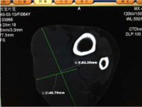
Figure 1
CT Solid mass infiltrated into the subcutaneous fat.
Figure 2
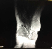
Figure 2
A MRI Signal intensity similar to the muscle on T1W1.
Figure 3
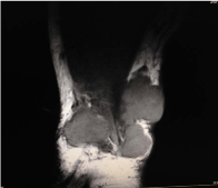
Figure 3
MRI Signal intensity slightly higher than that of muscle on T2W2.
Figure 4
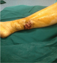
Figure 4
The tumor reappeared after one year later, a 6.0×7.0×15.0cm mass appeared on the incision.
Figure 5
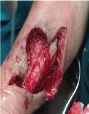
Figure 5
The resection of the mass. Part suture.
Figure 6
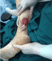
Figure 6
VSD covered the defect of the skin.
Figure 7
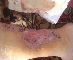
Figure 7
The tumor reappeared again after 10 days.
Figure 8
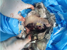
Figure 8
The tumor grew faster combined with necrosis, hemorrhage and foul smell in 3 months.
Figure 9
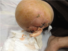
Figure 9
The amputation of the knee.
Figure 10
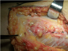
Figure 10
The new found tumor about 1.6.×1.6×1.0cm on the back of the knee.
Figure 11
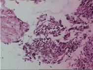
Figure 11
Tumor was composed of small, elongated cords or tubules, in a tightly packed arrangement (hematoxylin and eosin; magnification, ×10).
Figure 12
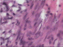
Figure 12
Tumor cells were smaller and cube-shaped or oval and low-grade
nuclei. (hematoxylin and eosin; magnification, ×40).
References
- Podetta M, D'Ambrosio G, Ferrari A, Sgarella A, Dal Bello B, Fossati GS, et al. Low-grade fibromatosis-like spindle cell metaplastic carcinoma: a basal-like tumor with a favorable clinical outcome: Report of two cases. Tumori. 2009; 95: 264-267.
- Sun N, Fu Y, Wang Y, Tian T, An W, Yuan T. Mucinous tubular and spindle cell carcinoma of the kidney: A case report and review of the literature. Oncl Lett. 2014; 7: 811-814.
- Velazquez EF, Werchniack AE, Granter SR. Desmoplastic/spindle cell squamous cell carcinoma of the skin. A diagnostically challenging tumor mimicking a scar: clinicopathologic and immunohistochemical study of 6 cases. Am J Dermatopathol. 2010; 32: 333-339.
- Fletcher CDM, Unni KK, Mertens F. World Health Organization of tumors. Pathology and genetics of tumors of soft tissue and bone. Lyon: IARC ress. 2002; 86-90.
- Gengler C, Guillou L. Solitary fibrous tumor and haema Pericy-toula evolution of a concept. Histopathology. 2006; 48: 63-74.