Review Article
The Management of Advanced Colorectal Carcinoma Having Failed Standard Therapy – An Immunologic Approach
Arlen M1*, Arlen P3,Saric O3, Dubeykovskiy A3, Coppa G1, Crawford JM2, Conte C1, and Molmenti E1
1Department of Surgery, Division of Surgical Oncology, North Shore University Hospital, USA
2Deptartment of Pathology, Division of Surgical Oncology, North Shore University Hospital, USA
3Division of Surgical Oncology, Northwell Health System, Hofstra College of Medicine and Precision Biologics Inc, USA
*Corresponding author: Myron Arlen, Division of Surgical Oncology, Department of Surgery North Shore University Hospital 450 Lakeville Rd. New Hyde Park NY. 11042, USA
Published: 10 Jul, 2016
Cite this article as: Arlen M, Arlen P, Saric O, Dubeykovskiy A, Coppa G, Crawford JM, et al. The Management of Advanced Colorectal Carcinoma Having Failed Standard Therapy – An Immunologic Approach. Clin Surg. 2016; 1: 1064.
Abstract
Over the past 40 years all of the major improvement in management of solid tumor malignancies including those of colorectal cancer has been via introduction of newer chemotherapeutic agents. During this time, while there have been significant changes in response as well as duration of survival, curative responses have not been noted. An obvious approach is to employ the patients own immune system to bring the developing lesion under control. It appears that while the malignancy does behave as a foreign cell type which when properly identified by host immunity can be controlled, the level of immunogenic protein characterizing the tumor is present at level far below what is immunologically required. Our group has defined these proteins from pooled allogeneic tumor membranes and delivered them at the threshold level felt to be required. Here, in early FDA approved clinical trials, significant enhancement in survival was accomplished. Considering that on completion of these early trials, the FDA considered that such future preparations were too dangerous, realizing that certain unrecognized viruses could be present. In order to more clearly define the antigen, monoclonals were developed for immunopurification, but on careful examination of the end results, it appeared that it was the antibody response via ADCC that initiated antitumor activity. The first of the mAbs related to the more prevalent oncofetal protein, MUC5ac was produced GMP for clinical trials.
Introduction
In order to control advanced colorectal cancer we are employing specific monoclonals antibodies
targeting oncofetal immunogenic proteins expressed by the tumor. We have completed Phase II
FDA mAb therapy trials with evidence of activity in heavily pretreated patients with metastatic
disease. Our group, utilizing pooled allogeneic tumor membrane protein obtained from operative
specimens post colectomy, has identified 3 oncofetal proteins that characterize the malignancy.
These proteins are not expressed in normal colon specimens adjacent to the tumor. The levels of
expression of such proteins are for the most part too low for the host to recognize the presence of
the lesion and as such are unable to turn on an effective immune response.
By pooling the tumor membrane proteins, the threshold level of antigen needed for acting
as a vaccine to turn on an immune response has been found to be at approximately 500 ugm
for the specimen with the tumor having been found to be expressing no more than 10-25 ugm
[1]. Progression follows once a lesion is established since with the initial appearance of matrix
metalloproteinase in the lesion this molecule not only helps with chaperoning the tumor cells to the
invading vascular supply, but by breaking down the e-cadherin to form small e-cadherin tending to
help in dissemination of the lesion [2]. Monoclonals targeting the small e-cadherin have been shown
to diminish progression of lesion spread and may be of eventual use in supporting our therapeutic
monoclonals.
While most believe that the major host response any immunogen is associated with, is primarily
a cytotoxic T cell reaction, we have clearly demonstrated that the anti tumor response is achieved via
IgG1 production with the initiation of Antibody Dependent Cell Cytotoxicity (ADCC) that activates
tumor destruction [3]. We have defined that there are 3 immunogenic proteins expressed in various
combinations in the colorectal lesion and similarly found to some
extent in pancreatic cancer (Figure 1) illustrates the need for a chimeric
or humanized form of IgG1 since the ADCC mechanism involves
delivery of the NK cell to the surface of the malignance lesion. It is
essential for a human Fc component of the IgG1 that contains the
NK cell receptor [4]. As such it is an antibody associated response
rather than direct activity of the monoclonal on the tumor. The mAbs
so derived from the existing oncofetal proteins appear to have an
extremely high targeting response to the existing lesion (ADCC) than
other antibodies associated with growth factors on the tumor, such
factors being present to some extent on normal tissue i.e. Anti-VEGF,
Avastin, Herceptin, and Erbitux.
Figure 1
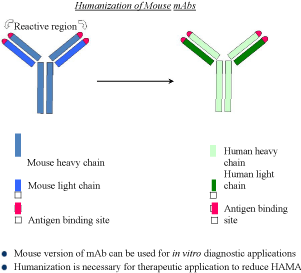
Figure 1
The presence of the human heavy chain contains receptors for the NK cell
Figure 2
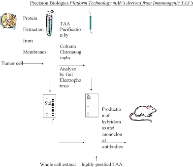
Figure 2
Illustration describes the presently employed Platform Technology. It illustrates the isolation of the immunogenic protein defining the presence of colon
cancer and its use in eventually producing those monoclonals capable of diagnosing and treating the targeted neoplasm.
Materials and Methods
The oncofetal proteins that we have identified as being expressed
in selected colorectal neoplasms demonstrate sequence homology to
CEACAM 5, 6, A33 and MUC5ac [5]. These molecule structures have
importance in the development of the fetus in the latter trimester.
Later they have their initiating genes re: methylated at term to
prevent further functionality. The oncofetal variants appeared to be
randomly expressed later in the development of the normal adult
tissue state, where either an oncogenic virus or carcinogen initiates
the transformation of the re: methylated gene resulting in appearance
of the carcinogenic effect [5]. Should the MUC5ac gene fail to be
re: methylated at birth then the result is the appearance of clinical
cystic fibrosis. The monoclonal antibody related to MUC5ac in fetal
tissue which is responsible for the production of mucin in the fetal
bowel or lung parenchyma will not react with the posttranslational
modification seen in the tumor. Neither will our NEO-102 antibody
respond to the normal MUC5ac antigen [6].
When present in the malignant lesion these oncofetal proteins
are again re-expressed, but at sub threshold levels preventing a host
response to the developing tumor system [7]. When the monoclonals
antibodies derived from these antigens are delivered at threshold
levels in their humanized or chimeric forms via the IV route they
attack those cells expressing the target protein in the suboptimal
tissue levels. One can see an effective immune response in bringing
the tumor under some degree of control beyond what can be
accomplished with other immunogens [8]. Vaccines using whole
cell preparations with a GVAX or Dendritic cell approach have not
been that effective in that they require suboptimal level of undefined
immunogen to be presented to the host at hopefully therapeutic titers.
In our studies, translating what has been found with animal models
receiving our antibody translates into relatively high doses of the mAb.
Approximately 400 mg/ of, monoclonal antibody are administered q
2 wks. To achieve what appears to be a potential clinical response [8].
The platform technology that Precision Biologics has followed is
illustrated in (Figure 2). It outlines the basics from which we have derived
our monoclonals and have moved on to tumor identification, the
later occurring by using affinity preparations for mass spectroscopy.
With the antigen and associated monoclonals at hand we were able
to show the effectiveness of the naked antibody in targeting nude
mice challenged with human colon and pancreatic tumor transplants
and to effectively demonstrate to the FDA the activity of the IgG1 in
suppressing tumor growth.
GMP preparations of monoclonal 101 further upgraded the
output employing a high expression vector system to produce Neo-
102. In (Figure 3) we see how changes in the system employed can
eventually lead to the therapeutic levels needed to come up with a
product for eventual commercial use (Figure 3) [3].
The trials employing the high production system were established
and have now completed the Phase II FDA studies and as reported
have shown indications of a significant benefit to the host. Since this
MUC5ac related antigen is the most common immunogen found
among the tumor specimens examined, we are specifically targeting
that protein, now termed Neo-102 with the corresponding mAb.
The study we performed as part of the clinical trial that defined
the sequence of the mutated MUC5ac protein and provided the
peptidomimetics of an NPC-1 epitope derived from MUC5AC. This
included composition comprising the amino acid sequence of the
epitope binding site defined by Phage display. That is the polypeptide:
F(PHE) P(PRO) E(GLU) D(ASP) Y(TYR) F(PHE) R(ARG) Y(TYR)
T(THN(ASN) Q((GLN) K(GLY). This sequence proved to be that
fragment of the 600kd MUC5ac molecule responsible for antibody
production and thus could potentially be utilized for eventual peptide
vaccine therapy as a preventative post surgical resection in high risk
patients.
In 2015 at the ASCO GI Symposium in San Francisco, CA , results
of the Phase 1 study of Ensituximab (Neo-102) in chemotherapy
refractory metastatic colorectal Cancer Study were presented in a
poster session. The study revealed a Maximal Tolerated Dose of
3.0 mg/kg IV every 2 weeks. The overall survival observed in this
demonstrated 10.4 months comparing favorably to the historical
control for a similar population of patients with advanced colorectal
Ca (5 months). This led to a larger Phase 2 multi-center colorectal
cancer study using Ensituximab in the same patient population.
In examining a higher powered view of the colon cancer
specimen defined in (Figure 4), at 20 power it is apparent that those
cells at the margin of the intestinal villiare expressing tumor protein
which was expected to be shed into the lumen of the bowel. We have
developed a simple office stool ELISA test to exam the stool of a
patient for diagnostic purposes. When we can show the presence of
a small amount of antigen present in the stool, it would represent
for purposes, the presence of an early intraepithelial lesion unrelated
to polyp developing within a field effect of the bowel [9]. Absence
of antigen would signify that the bowel is free of any developing
malignant lesion and that colonoscopy can be delayed.
Figure 3
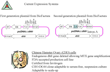
Figure 3
Illustrating those modifications present in the first and second plasma vector.
Figure 4
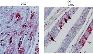
Figure 4
In a careful evaluation of the Immunohistochemical analysis of those cells express 2 discrete tumor antigens that are 31.1 and Neo 102 it is apparent that
only those cells representing the malignancy will be identified on IHC
Conclusion
In evaluating the initial FDA phase II studies to date it appears that the initial premise that immunotherapy has advantages in controlling advanced and metastatic colorectal cancer does hold true. To the best of our knowledge, there is no data to date that suggests that any patient with a solid tumor malignancy that has metastasized has been cured by chemotherapy alone. The data obtained from this initial monoclonal trial is leading to a planned for clinical Phase III study where low dose chemotherapy will be added to remove inhibitory blocking substances shown to be expressed by the tumor Eventually additional therapeutic modalities will be added to the chemoimmunotherapy planned for trial. Here IL-15 will be considered for use in enhancing ADCC and eventually a radiolabelled alpha emitter of Neo-102 will also be utilized to enhance the overall response. We anticipate that this approach should markedly improve the results that we are able to obtain by the use of chemotherapy alone.
References
- Elias EG, Hollinshead A, Arlen M, Mosely MS. Adjuvant-Specific Active Immunotherapy in Patients with Colon Adenocarcinoma Utilizing Polypeptide Tumor Associated Antigens (TAA): Proceedings Am Soc Clin Oncol. Oncol. 1985; V-4, 76.
- Tsang KY, Johnson E, Bishop L, LaVia, Warren RQ, Christian M, et al. Monoclonal Antibodies to Human Colon Carcinoma-Associated Antigens .Abst. Cancer Detection and Prevention. International Soc. Preventive Oncology. 1987; 11: 1/2.
- Hollinshead AC, Stewart THM, Elias G, Arlen M. Co-assesment of Serum Epitope Antibodies,Cell Mediated Immunity and Survival in Colon Cancer Patients on TAA Specific Active Immunotherapy. Proc. Sixth Int. Conf. on Adjuvant Therapy of Cancer. W.B.Saunders. Section IX Colorectal Cancer, 1990; 454-468.
- Arlen M, Tsang KY. The Nature of the Monoclonal Antibodies Derived from Immunogenic Membrane Antigen of Human Colon Carcinoma Origin. J Tumor Marker Oncology. 1990; 5: 313-319.
- Arlen M, Arlen P, Bristol A, Luka J, Kantor J, Wang X. The Dual Functionality of Monoclonal Antibodies Derived from Tumor Associated Antigens. J Surgical Oncology. 2010.
- Arlen M, Arlen P, Tsang A, Wang XP. The Therapeutic Value of Monoclonal Antibodies Directed Against Immunogenie Tumor Glyproteins J Cancer. 2010; 1: 209-222.
- Hollinshead A, Elias G, Arlen M, Mosely MS, Scherrer J. Specific Active Immunotherapy in Patients with Adenocarcinoma of the colon utilizing TAA – Cancer. 1985; 56: 480-489.
- Arlen M, Arlen P. Optimizing the Immune System to Achieve Control of the Metastatic Malignant Lesion. J Cancer. 2013; 4: 427-432.
- Arlen M, Saric O, Wang X, Dubeykonskiy A, Arlen P. Nannocytology vs. Immunohistochemistry of Intestinal Colonocytes to Assess the risk of Colon Cancer Based on Field Cancerization. J Cancer. 2013; 4: 165-169.