Case Report
Keratocystic Odontogenic Tumor Mimicking Lateral Periodontal Cyst: A Case Report
Alparslan Esen1*, Ali Kilinc1, Yigit Guler1, Melek Tassoker2 and Omer Gunhan3
1Department of Oral and Maxillofacial Surgery, University of NecmettinErbakan, Turkey
2Department of Oral and Maxillofacial Radiology, University of NecmettinErbakan, Turkey
3Department of Pathology, Gulhane Military Medical Faculty, Turkey
*Corresponding author: Alparslan Esen, Department of Oral and Maxillofacial Surgery, NecmettinErbakan University, KaraciganMh. Ankara Cd. No: 74/A, 42050 Karatay, Konya, Turkey
Published: 15 Jul, 2016
Cite this article as: Esen A, Kilinc A, Guler Y, Tassoker M, Gunhan O. Keratocystic Odontogenic
Tumor Mimicking Lateral Periodontal Cyst: A Case Report. Clin Surg. 2016; 1: 1045.
Abstract
The aim of this case report is to present a keratocystic odontogenic tumor (KCOT) mimicking
lateral periodontal cyst. A 45-year-old female patient showed a well-defined radiolucent lesion
between the roots of the left second premolar and left first molar. The clinical and radiographic
features lead us to diagnose a lateral periodontal cyst that was treated by enucleation with curettage.
Histological evaluation showed typical histological features of KCOT. The clinical findings and the
radiological features are not distinctive enough to give a diagnosis of any bone lesion occurring in
the maxillofacial area. Therefore, histopathological evaluation is required for definitive diagnosis.
Keywords: Keratocystic odontogenic tumor; Lateral periodontal cyst
Introduction
Odontogenic keratocyst’s (OKCs) are benign cysts usually affecting the posterior mandible
which were first described by Phillipsen in 1956 [1]. According to latest World Health Organization
classification, OKCs were termed as keratocystic Odontogenic Tumors (KCOTs) because of its
neoplastic nature and tumor like characteristics of the lining epithelium. KCOTs differ from
other odontogenic cysts because of its aggressive behavior, specific histopathological features and
tendency to recurrence. Moreover it may be associated with Nevoid Basal Cell Carcinoma Syndrome
or Gorlin Syndrome [2].
The most common clinical symptoms of KCOTs are swelling, pain and discharge but it may
also be asymptomatic. The mandible is affected more than maxilla. It is commonly seen in ramus,
third molar area and body of mandible respectively. KCOTs appear more in males than females and
male to female ratio is 1.6:1. It may affects patients in a wide age range especially in second and third
decade [2,3].
Radiographically, KCOTs usually appear unilocular asymptomatic radiolucency with
well-defined sclerotic borders or multilocular radiolucency. Unilocular KCOTs can mimic
radiographically dentigerous cysts by surrounding crown of unerupted teeth, lateral periodontal
cysts by locating between the roots of teeth or nasopalatine duct cyst by locating maxillary midline
[4].
Lateral periodontal cysts (LPCs) are defined as non-keratinized developmental cysts located
adjacent or lateral to the root of vital teeth [5]. The diagnosis of lateral periodontal cysts is primarily
based on histopathologic features, as certain characteristic histologic features separate it from other
odontogenic cysts [6]. The aim of this case report was to present a new case of KCOT mimicking
radiographically LPC.
Case Report
A 45-year-old female patient was referred to the oral and maxillofacial surgery clinic in April 2015 with a 1-month history of painless swelling in the posterior mandible. The patient did not any systemic disease, and intraoral examination revealed a swelling on the vestibular side of the second premolar and the first molar (Figure 1). The teeth were found to be vital to electric pulp testing. In the radiologic examination, a well-defined radiolucency was detected between the roots of the second premolar and first molar on a panoramic radiograph (Figure 2). Aspiration revealed pale yellow straw colored fluid. Lateral periodontal cyst was considered in the clinical diagnosis because of the localization of the lesion and vital teeth. An excisional biopsy was performed under local anesthesia (Figure 3). The lesion was completely enucleated with a curettage and mucoperiosteal flap was sutured (Figure 4 and 5). Histologically, KCOT was diagnosed. A parakeratinized keratocystic wall, with variable thickness, overlying a fibrous stroma was observed in the microscopic section. Typical palisading basal cells of KCOT and luminal keratin formation were conspicuous. Focal suprabasal dysplasia and subepithelial hyalinization were observed (Figure 6). The patient was examined for clinical and radiologic follow up at 1, 3, 6 and 12 months. Complete recovery was seen after 12 months radiographically (Figure 7).
Figure 1
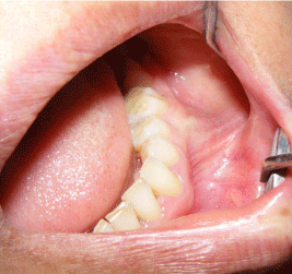
Figure 1
Intraoperative view of the lesion.
Figure 2
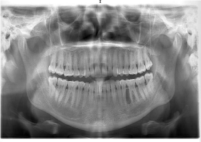
Figure 2
Preoperative radiographic view of the case. A well-demarcated
radiolucency is seen on the left posterior mandible.
Figure 3
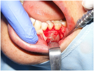
Figure 3
Intraoperative view of the lesion.
Figure 4
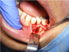
Figure 4
Intraoperative view of the lesion after enucleation with curettage.
Discussion
KCOTs are considered to be developmental cyst which arise from
the dental lamina remnants or primordial tissues like enamel organ.
These tumors are often seen in the mandible, especially in the posterior
body and ramus regions and it can cause the migration of the adjacent
teeth. They are usually asymptomatic. However, if the mass closer to
the anatomical structures, patients can suffer from pain or paresthesia
in the lower lip [7,8]. In this case, the lesion was observed between the
roots of the teeth in the molar region as unusual. The patient did not
complain of pain or paresthesia. But she reported to us with a chief
complaint of mild swelling.
Even if the radiographic images of the maxillofacial lesions which
are formed in the bone give an idea for the diagnosis, histological
examination is required for definitive diagnosis. In addition, the
aspiration punctures as a tool can also give an idea for the early
diagnosis. Generally, dirty, creamy white and cheese like materials
can be obtained from the aspiration fluid in KCOTs [9]. In this
case, aspiration revealed pale yellow straw colored fluid. Hence, we
considered that the lesion would be a lateral periodontal cyst because
of the reasons including radiographic appearance, color of aspiration
fluid and the vital teeth adjacent to the lesion. However, excisional
biopsy showed that the mass was a KCOT.
LPCs are developmental odontogenic cysts and it can be
clearly differentiated from KCOT histopathologically. LPC is lined
by thin, non-keratinized epithelium usually of one to five layers
thick. However, KCOTs are usually lined by parakeratinized or
orthokeratinized epithelium which is highly characteristic with 6-10
layers ridges [2,10]. In this case, parakeratinized epithelium was
observed histologically.
The recurrence rate of the KCOTs varies between 2.5% and 62.5%
[11]. The parakeratotic type of KOCTs has a high recurrence rate and
locally destructive character. Most recurrences take place within 5–7
years after surgery, although some have been reported more than 10
years following initial intervention [12]. The histological evaluation
of parakeratotic type shows a regular parakeratinized stratified
squamous epithelium with a corrugated surface and palisaded basal
cells. These findings emphasize the importance of long-term followup
of KCOTs. In our case, we thought possibility of recurrence
because of the parakeratinized epithelium. The patient was followedup
periodically and the completely bone regeneration was observed
on panoramic radiograph 12 months after surgery. However, the
patient is still under our control.
There are many methods used for the treatment of KOCTs.
The surgical treatment methods are categorized by Morgan et al.
[12] as conservative or aggressive. Conservative treatment includes
enucleation, with or without curettage, or marsupialization. Its
advantage is preservation of anatomical structures (including teeth)
especially in young patients. Aggressive treatment includes peripheral
ostectomy, chemical curettage with Carnoy’s solution or en bloc
resection. Aggressive modalities have generally been recommended
for nevoid basal cell carcinoma cases, large KCOTs and recurrent
lesions [13]. In this case we preferred enucleation with curettage.
In the literature, there are a few case reports of keratocystic
odontogenic tumor mimicking lateral periodontal cyst. Hiremath
et al. [14] reported a case report of a 45 year old woman with an
asymptomatic swelling on the gingiva between the mandibular left
premolars. They concluded that the pathologic specimens should be
submitted for histopathological evaluation for a definitive diagnosis
due to high recurrence rate and aggressive behavior of OKCs.
However, they did not give information about the follow-up period
for their case. Borgonovo et al. [15] stated that a 32-year-old female
patient showed a well-delimited radiolucent lesion connected with the
root of the left third molar with close anatomical relationship with the
mandibular canal. They reported that the lesion showed radiologically
complete remission without any recurrence 1 year after surgery.
Bojan et al. [16] also reported a case of OKC situated in between the
roots of lower right premolars resembling LPC. They informed that
the patient was completely asymptomatic after 1 month follow-up
postoperatively. As known, KCOTs are characterized by a benign but
locally invasive behavior with a high risk of recurrence. KCOTs can
spread to adjacent soft tissues and it can show infiltration into the
bone, and destructive growth [11,12]. Some immunohistochemical
studies suggest that this bone destruction may be associated with
the osteoclastogenesis [17-19]. Bone resorption regulators play an
important role in bone resorption activity. The Receptor Activator
of Nuclear Factor kappa B (RANK) belongs to the Ttumor Necrosis
Factor (TNF) receptor super family and is activated by RANK ligand
(RANKL). RANKL is a homotrimetric protein and it activated T
cells. RANKL binds to RANK on the surface of pre-osteoclasts
and stimulates the development and activation of osteoclasts.
Osteoprotegerin (OPG) interrupts this activation by binding directly
to RANKL. Therefore, bone resorption and formation are regulated
by RANK-RANKL and OPG levels [20]. Da silva et al. [17] examined
these bone resorption regulators by using immunohistochemistry in
the odontogenic tumors and they demonstrated differences in the
expression of these molecules in odontogenic epithelial tumors. They
emphasized that the imbalance of these factors could contribute to
the differential bone/tooth resorption activity in odontogenic lesions.
Andrade et al. [18] investigated the expression of the same bone
resorption regulators in odontogenic tumors and they reported that
the RANKL/OPG ratio was different in various odontogenic tumors.
Tekkesin et al. [19] have found that a greater number of RANK-positive
cells in the epithelial component of OKCs and ameloblastomas than
in radicular cysts. They reported that the molecular system of RANK/
RANKL/OPG is variably expressed in odontogenic lesions and this
system may be involved in the osteoclastogenic mechanisms in OKCs
and ameloblastomas.
In conclusion, the maxillofacial bone lesions can resemble
each other radiographically. Moreover, they can have similar
clinical symptoms. Therefore, we considered that histopathological
examination should be performed for a definitive diagnosis for all
bone lesions.
Figure 5
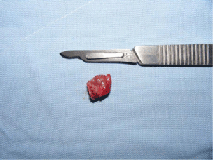
Figure 5
Biopsy material.
Figure 6
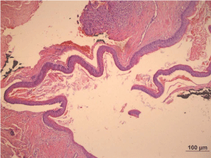
Figure 6
Figure 6 shows a parakeratinized keratocystic wall, with variable
thickness, overlying a fibrous stroma. Typical palisading basal cells of KCOT
and luminal keratin formation are conspicuous. Focal suprabasal dysplasia
and subepithelial hyalinization are observed. (H&E stained, Original
magnification:100x).
Figure 7
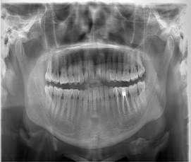
Figure 7
Complete healing was seen after 12 months.
References
- Philipsen HP. Om keratocystedr (Kolesteratomer) andkaeberne. Tandlaegebladet. 1956; 60: 963–971.
- Grasmuck EA, Nelson BL. Keratocystic odontogenic tumor. Head Neck Pathol. 2010; 4: 94-96.
- Shear M. The aggressive nature of the odontogenic keratocyst: is it a benign cystic neoplasm? Part 2. Proliferation and genetic studies. Oral Oncol. 2002; 38: 323-331.
- MacDonald, Jankowski DS. Keratocystic odontogenic tumour: systematic review. Dentomaxillofac Radiol. 2011; 40: 1-23.
- Formoso Senande MF, Figueiredo R, Berini Aytés L, Gay Escoda C. Lateral periodontal cysts: a retrospective study of 11 cases. Med Oral Patol Oral Cir Bucal. 2008; 13: E313-317.
- Mendes RA, van der Waal I. An unusual clinicoradiographic presentation of a lateral periodontal cyst--report of two cases. Med Oral Patol Oral Cir Bucal. 2006; 11: E185-187.
- Regezi JA, Sciubba JJ, Jordan RCK. Oral Pathology: Clinical Pathologic Correlations. Elsevier Health Sciences. 2012.
- Bande CR, Prashant MC, Sumbh B, Pandilwar PK. Prevalence, treatment and recurrence of odontogenic keratocyst in central India. J Maxillofac Oral Surg. 2010; 9: 146-149.
- Purkait SK. Essentials of Oral Pathology. 2011: Jaypee Brothers, Medical Publishers Pvt. Limited.
- Sulijak JP, Bohay RN, Wysocki GP. Lateral periodontal cyst: a case report and review of the literature. Can Dent Assoc. 1998; 64: 48-51.
- Forssell K, Forssell H, Kahnberg KE. Recurrence of keratocysts. A longterm follow-up study. Int J Oral Maxillofac Surg. 1988; 17: 25-28.
- Morgan TA, Burton CC, Qian F. A retrospective review of treatment of the odontogenic keratocyst. J Oral Maxillofac Surg. 2005; 63: 635-639.
- Madras J, Lapointe H. Keratocystic odontogenic tumour: reclassification of the odontogenic keratocyst from cyst to tumour. J Can Dent Assoc. 2008; 74: 165-165h.
- Hiremath SS, Deshpande AM, Byakodi S, Magdum DB. Diagnostic Dilemma: A Case Report of Odontogenic Keratocyst in Lateral Periodontal Position. Int J Oral Maxillofac Pathol. 2011; 2: 23-26.
- Borgonovo AE, Bernardini L, Francinetti P, Rizza F, Re D. Odontogenic keratocyst mimicking paradental cyst. Case Rep Dent. 2014; 2014: 974241.
- Bojan A, Duraiselvi P, Sumathy C, Mithra. Odontogenic Keratocyst Simulating Lateral Periodontal Cyst: A Case Report. Int J Adv Health Sci. 2015; 2: 1-4.
- da Silva TA, Batista AC, Mendonça EF, Leles CR, Fukada S, Cunha FQ. Comparative expression of RANK, RANKL, and OPG in keratocystic odontogenic tumors, ameloblastomas, and dentigerous cysts. Oral Surg Oral Med Oral Pathol Oral Radiol Endod. 2008; 105: 333-341.
- Andrade FR, Sousa DP, Mendonça EF, Silva TA, Lara VS, Batista AC. Expression of bone resorption regulators (RANK, RANKL, and OPG) in odontogenic tumors. Oral Surg Oral Med Oral Pathol Oral Radiol Endod. 2008; 106: 548-555.
- Tekkesin MS, Mutlu S, Olgac V. The role of RANK/RANKL/OPG signalling pathways in osteoclastogenesis in odontogenic keratocysts, radicular cysts, and ameloblastomas. Head Neck Pathol. 2011; 5: 248-253.
- Moraesa M, Matosa FR, Lopes Costa AL. Osteoclastogenesis regulatory factors (RANK, RANKL and OPG)in osteolytic jaw lesions. Rev OdontoCienc. 2013; 28: 53-57.