Case Report
Self-inserted Sewing Needles Through the Abdominal Wall: Is the Laparoscopic Approach with Omentectomy a Feasible Option?
Aktimur R1*, Cetinkunar S2and Kucuk OG3
1Department of General Surgery, Istanbul Aydin University, Turkey
2Department of General Surgery, Adana Numune Training and Research Hospital, Turkey
3Department of General Surgery, Samsun Training and Research Hospital, Turkey
*Corresponding author: Recep Aktimur, Department of General Surgery, Istanbul Aydin University, Istanbul, Turkey
Published: 09 Jun 2016
Cite this article as: Aktimur R, Cetinkunar S, Kucuk OG.
Self-inserted Sewing Needles Through
the Abdominal Wall: Is the Laparoscopic
Approach with Omentectomy a Feasible
Option?. Clin Surg. 2016; 1: 1029.
Abstract
Background: Ingestion of sewing needles is fairly common in daily emergency medical practice, but repetitive self-insertion of sewing needles through the abdominal wall is a rare condition.
Objective: This case is illustrated to point that in patients with self-insertion of a foreign body through the abdominal wall, laparoscopic approach should be considered with the advantage of magnifying effect and improved postoperative clinical outcomes.
Case:2In the present case, one subcutaneous and two intraperitoneal, non-ingested, forcefully inserted sewing needles were managed laparoscopically. The clinical presentation and laparoscopic technique were discussed under a review of recent literature.
Conclusion: Laparoscopic exploration has the advantage of magnified sight, which can be helpful in finding non-localized, intra-abdominal foreign bodies, when the object is stuck in the omentum.
Keywords: Foreign body; Sewing needles; Laparoscopy
Introduction
There are several intraperitoneal foreign bodies and a wide spectrum of presentations reported in the literature [1]. Foreign bodies may reach the intraperitoneal space via mouth, anus, urogenital tract, or percutaneously. This situation usually occurs accidentally or, rarely, as a result of deliberate self-harm. The majority of such cases occurs in children and mentally challenged patients. Clinical presentation can vary from asymptomatic to acute abdominal pain with an intra-abdominal abscess [2]. We present the case of a mentally unsound woman with one subcutaneous and two intraperitoneal foreign bodies as a result of deliberate self-harm. The laparoscopic approach for non-localized intraperitoneal sewing needle was discussed.
Case Presentation
A 42-year-old woman with known psychiatric illness was referred to our Emergency Department with symptomatic intra-abdominal foreign bodies. Her past medical history revealed several admissions to the emergency department with repetitive minor deliberate self-harm and she had undergone an appendectomy operation performed 4 years ago. She stated that she had acute onset abdominal pain for approximately a 24-hour period. Physical examination showed no peritoneal irritation, but a palpable foreign body was present at the described area. Laboratory findings and bowel movements were normal. The abdominal x-ray demonstrated three thin, radiopaque objects in the superior pelvic area (Figure 1A). Since she was complaining of pain, the decision was made to attempt a diagnostic laparoscopy and if it is possible a laparoscopic foreign body retrieval. To identify the exact location of the foreign bodies, a computed tomography (CT) was performed, which showed one subcutaneous and two intraperitoneal items of linear density (Figure 1B). The patient was informed of these findings.
To easily reach the two intraperitoneal foreign bodies, two 10 mm and one 5 mm trocar were
atypically placed (Figure 2A). A 300
10 mm camera was used. The intraperitoneal, infra-umbilical,
vertically placed sewing needle was easily removed, and then the intraperitoneal space was explored.
To identify the definite location of the transverse-placed object, intra-operative fluoroscopy was
performed. During the fluoroscopic exam, movement of the
radiopaque object was detected when the omentum was moved with
the laparoscopic instrument. The omentum was carefully examined,
but no existing foreign body was discovered. To avoid hemorrhage,
the blunt dissection was stopped and the decision was made to
perform an omentectomy. During this procedure, a sewing needle,
which was stuck in the omentum, was recognized and removed. The
omentectomy specimen was extracted from the infra-umbilical trocar
incision. Following extraction of the intraperitoneal needles, with
the use of laparoscopic camera light illumination, a subcutaneously
localized sewing needle was found and removed through a 2 cm
skin incision (Figure 2A and B). The postoperative course was
uneventful and the patient was discharged on postoperative day 2. No
complications or complaints were observed by the 1 month follow-up
examination.
Figure 1
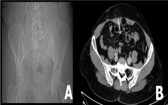
Figure 1
(A) X-ray image showing three thin radiopaque objects. (B) CT
showing the vertical intraperitoneal sewing needle.
Discussion
Ingested foreign bodies easily transit through the gastrointestinal tract. Foreign bodies localized in the digestive tract may pass uneventfully with feces within 4–6 days. But, predicting the route and location of an intraperitoneal needle is difficult. In less than 1% of patients does the sharp object perforate the gastrointestinal wall and migrate to the intraperitoneal area, or rarely a sharp object can cause an impalement injury in the intraabdominal organs through the abdominal wall [2,3]. Also, there are two published cases about the intraperitoneal migration of accidentally inserted sewing needles: one penetrated to the stomach and diaphragm in a 10-month-old male infant through the posterior abdominal wall, and the other penetrated through the lateral abdominal wall of a 13-year-old boy without any organ injury [4,5]. A literature review has shown that, self-ingested intraperitoneal foreign body cases have an incidental route, and two of three published cases described an associated organ injury. Additionally, non-ingested intraperitoneal foreign bodies may result in chronic abdominal pain. In the first assessment of our case, she was denied that any patient-related cause, but when her past medical history was taken into account, deliberate self-harm was suspected. In the second assessment, when she was informed about her radiological evaluation and subcutaneous palpable foreign body, she was accepted repetitive needle insertions through the abdominal wall. In this case no visceral organ injury was noted.
Figure 2
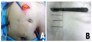
Figure 2
(A) Trocar placement and subcutaneous sewing needle removal
incision with the needle showing on the tip of the surgical instrument. (B) The
removed needles.
In a case of not-localized intraperitoneal foreign body, the patient can be an appropriate candidate for open surgery, or in selected patients a laparoscopic approach may be preferable [5,6]. In the present case, one subcutaneous and two intraperitoneal, non-ingested, forcefully inserted and not-precisely localized sewing needles were managed laparoscopically and through local exploration of the skin. This case is interesting by, no intra-abdominal organ injury despite the migration route, mechanism and used technique to facilitate needle removal. Sociocultural factors or co-existing psychiatric disorders of the patients may result in different clinical manifestations and this may result in some challenges in daily emergency practice. In our opinion, appropriate radiological evaluation guided laparoscopic exploration has the advantage of magnified sight, which can be helpful in finding non-localized, intra-abdominal foreign bodies, and when the object is stuck in the momentum, an omentectomy can be a safe and feasible option under fluoroscopic examination.
References
- Yalçin S, Karnak I, Ciftci AO, Senocak ME, Tanyel FC, Büyükpamukçu N. Foreign body ingestion in children: an analysis of pediatric surgical practice. Pediatr Surg Int. 2007; 23: 755-761.
- Maleki M, Evans WE. Foreign-body perforation of the intestinal tract. Report of 12 cases and review of the literature. Arch Surg. 1970; 101: 475- 477.
- Crawford DL, McVay WB. Delayed presentation of colonic impalement injury by picture frame glass fragment treated using hand-assisted laparoscopic colectomy. Surg Laparosc Endosc Percutan Tech 2008; 18: 619-621.
- Yalçin S, Karnak I, Ciftci AO, Senocak ME. An unusual penetrating injury in an infant: straight-pin migration from the back to the stomach. J Pediatr Surg. 2006; 41: 1332-1334.
- Aarabi S, Stephenson J, Christie DL, Javid PJ. Noningested intraperitoneal foreign body causing chronic abdominal pain: a role for laparoscopy in the diagnosis. J Pediatr Surg 2012; 47: 15-17.
- GRodriguez-Hermosa JI, Codina-Cazador A, Sirvent JM, Martin A, Girones J, Garsot E. Surgically treated perforations of the gastrointestinal tract caused by ingested foreign bodies. Colorectal Dis 2008; 10: 701-707.